Figure 4. Elevated canonical Wnt signaling in flow-sorted retinoblastoma cancer stem-like cells. A: Weri cells were sorted by flow cytometry using an antibody against the stem cell marker ABCG2. RNA and protein were then
extracted from the ABCG2-positive and ABCG2-negative cells. Western blotting on sorted ABCG2+ stem cells showed higher β-catenin
levels, indicating higher canonical Wnt signaling in the stem cells (n=3, p=0.048). β-catenin levels were normalized to β-actin,
and are expressed as a ratio of the respective ABCG2-negative cells. B-F: Representative images showing the immunoreactivity of the stem cell markers ABCG2 and Msi1, and the canonical Wnt signaling
mediator β-catenin in the retinoblastoma cell lines Weri and Y79. In the top right panels, ABCG2 immunoreactivity is red and
β-catenin is green. In the top left panels, Msi1 immunoreactivity is green and β-catenin is red. The bottom panels show the
negative controls, in which Weri and Y79 cells were treated with control rabbit IgG and FITC or TRITC secondary antibodies.
The adjacent bright-field images are also shown. All images were taken at 40x. G: QPCR on canonical Wnt signaling receptors demonstrated higher expression of the receptors LRP6, Fz3, Fz4, and Fz6 in the
in ABCG2-positive (cancer stem-like cells) than ABCG2-negative (non-stem-like cells). Data are from two independent experiments;
each experiment had three replicates.
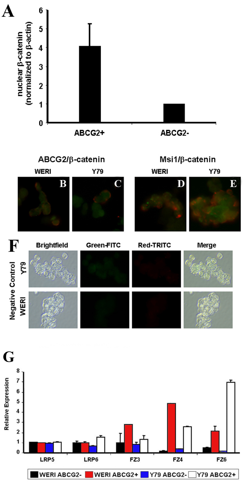
 Figure 4 of
Silva, Mol Vis 2010; 16:36-45.
Figure 4 of
Silva, Mol Vis 2010; 16:36-45.