Figure 4. Fundus photographs of patients
with mutations of TRPM1. A-C: Fundus images of patient
#373 (A), patient #437 (B), and patient #484 (C).
All fundus examinations revealed no abnormalities in the posterior pole
other than myopic changes, including the tilted discs and temporal
pallor of the optic discs. The patient’s number is indicated in each
photograph.
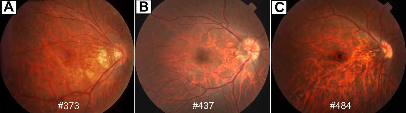
 Figure 4 of Nakamura, Mol Vis 2010; 16:425-437.