Figure 1. Compound heterozygous TRPM1
mutations identified in patients #373, #437, and #484. A:
Sequence chromatograms showing the mutations: IVS2–3C>G in patient
#373 (a), IVS8+3_6delAAGT in patient #484 (b), Arg624Cys (c.
1870C>T) in patients #437 and #484 (c), Ser882Ter (c. 2645C>A) in
patient #437 (d), and Phe1075Ser (c. 3224T>C) in patient #373(e). B:
Complete
CSNB pedigrees of Japanese patients #373, #437, and #484.
These three patients are compound heterozygotes of TRPM1
mutations. C: Exon structures of human TRPM1. The first
methionine (Start) and a stop codon (Stop) of the TRPM1 open
reading frame are indicated. All mutations found in this study are
shown. D: Putative topology of the human TRPM1. All mutations
found in this study are illustrated. The six transmembrane domains are
indicated as TM1-TM6. E: Alignment of R624 and F1075 in TRPM
proteins. Sequence alignment of TRPM1 from human, bovine, mouse, chick,
and TRPM2, TRPM4, and TRPM7 from human. Amino acid residues R624 and
F1075 are boxed. The asterisks indicate completely conserved residues.
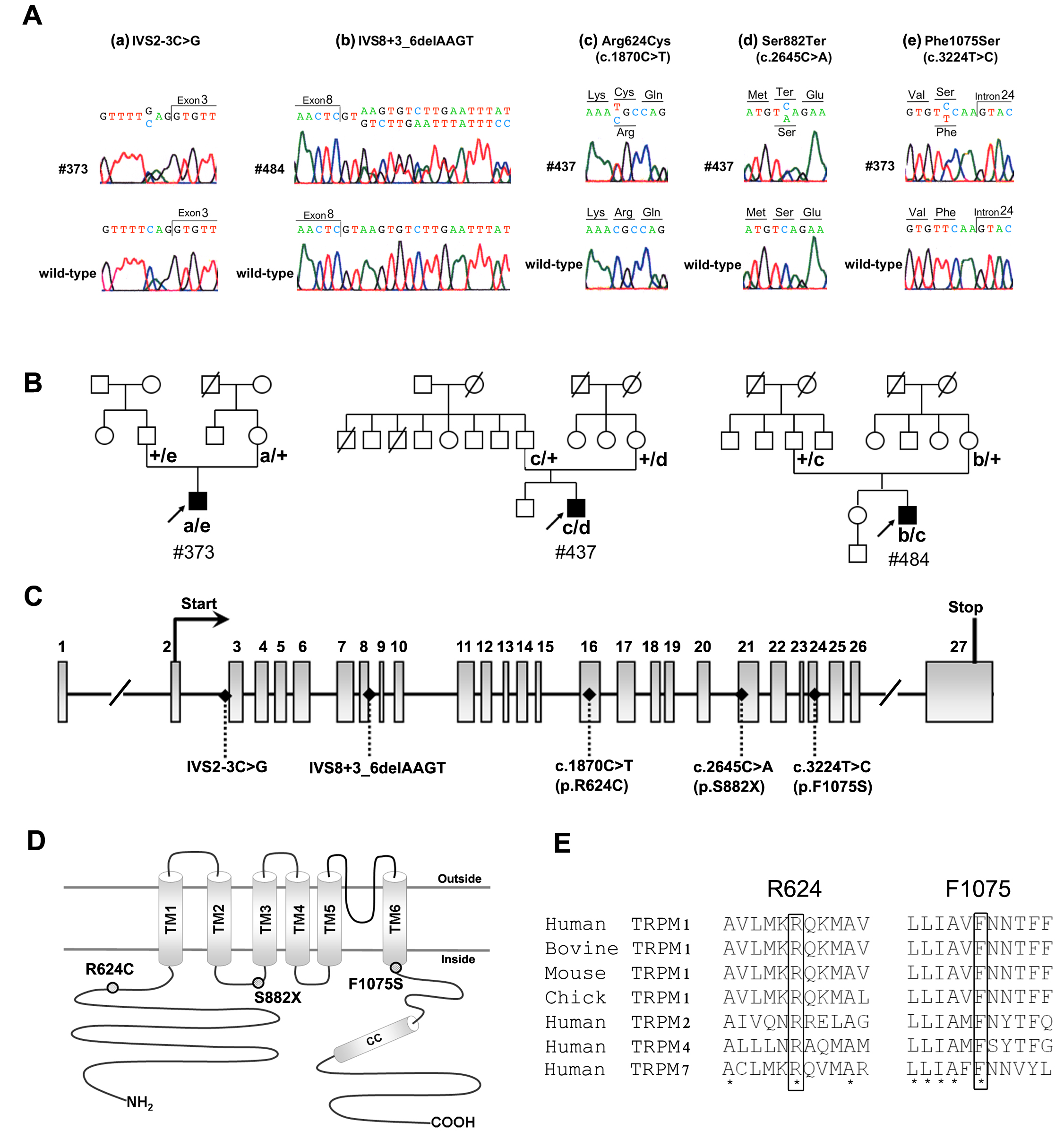
 Figure 1 of Nakamura, Mol Vis 2010; 16:425-437.