Figure 7. Phosphorylated p38
immunofluorescence in the retinal arteries and neuroretina. A:
A representative example of phosphorylated p38 (p-p38; green) staining
of the retinal arteries is shown. Double staining with the smooth
muscle cell marker smooth muscle actin (SMA; red) and the endothelium
cell marker CD31 (red) shows that p-p38 is primarily located in the
smooth muscle layer. Similar results were seen for sham-operated and
ischemia-reperfusion eyes. B: p-p38 (green) was also
occasionally detected in the inner nuclear layer of the neuroretina.
Double staining with protein kinase Cα (PKCα; red), a bipolar cell
marker, showed that p38 was localized to bipolar cell bodies, whereas
the p38 protein appears to be associated with the nucleus. The
abbreviations used in the figure are outer nuclear layer (ONL), outer
plexiform layer (OPL), and inner plexiform layer (IPL).
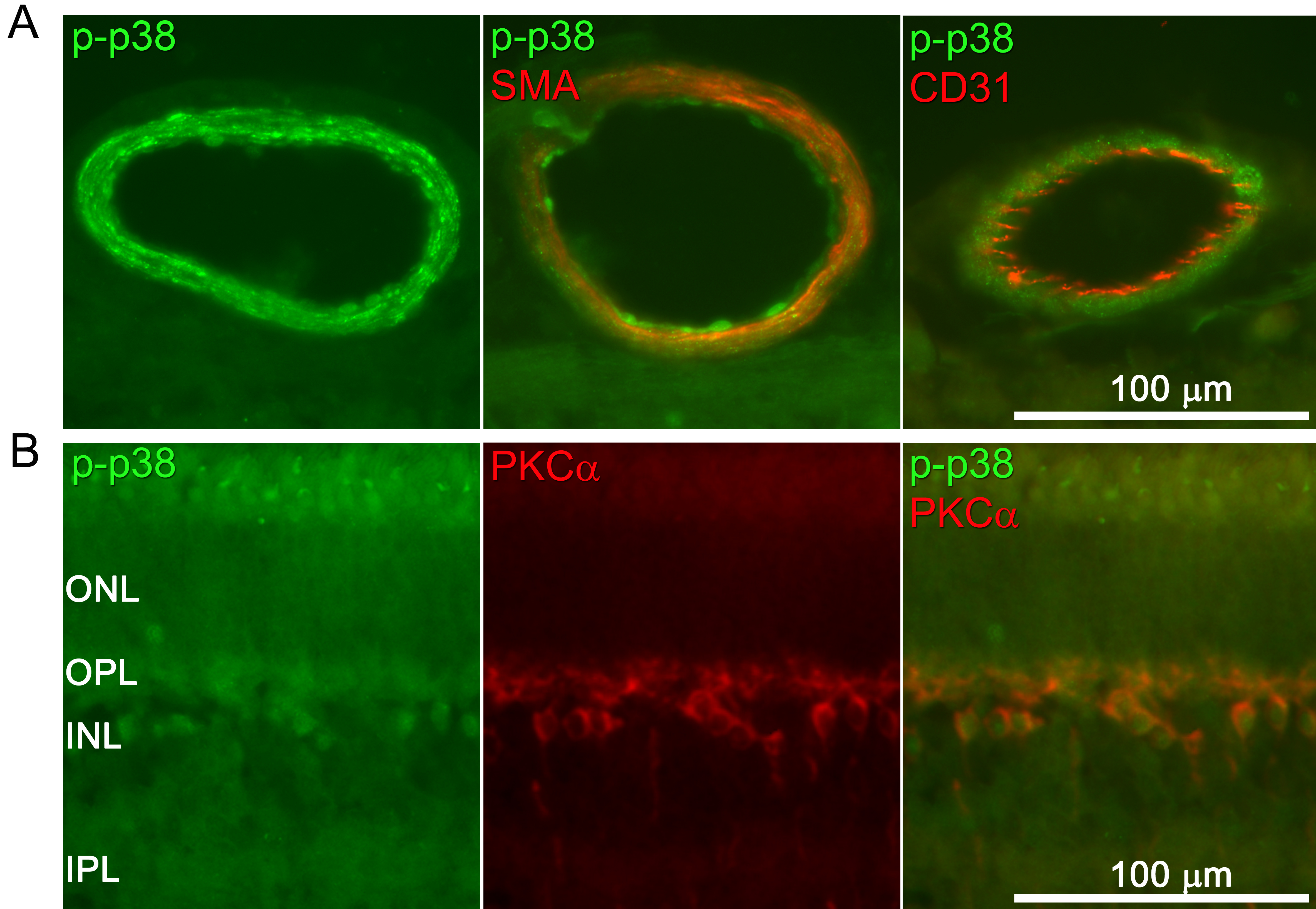
 Figure 7 of Gesslein, Mol Vis 2010; 16:392-407.