Figure 4. Phosphorylated extracellular
signal-regulated kinase 1 and 2 (ERK1/2) immunofluorescence in the
retina. Immunofluorescence staining of retina from pigs subjected to
ischemia followed by 5 (n=4), 12 (n=5), or 20 (n=4) h of reperfusion
and the corresponding sham-operated eyes. Note the enhanced staining
for phosphorylated ERK1/2 following retinal ischemia, especially after
5 h of reperfusion. Immunofluorescence staining for ERK1/2 appears to
be located in the Müller cells, including both the cell bodies and in
the radial processes, and in the inner retina, probably corresponding
to Müller cell endfeet and astrocytes. The abbreviations used in the
figure are outer nuclear layer (ONL), inner nuclear layer (INL), and
ganglion cell layer (GCL).
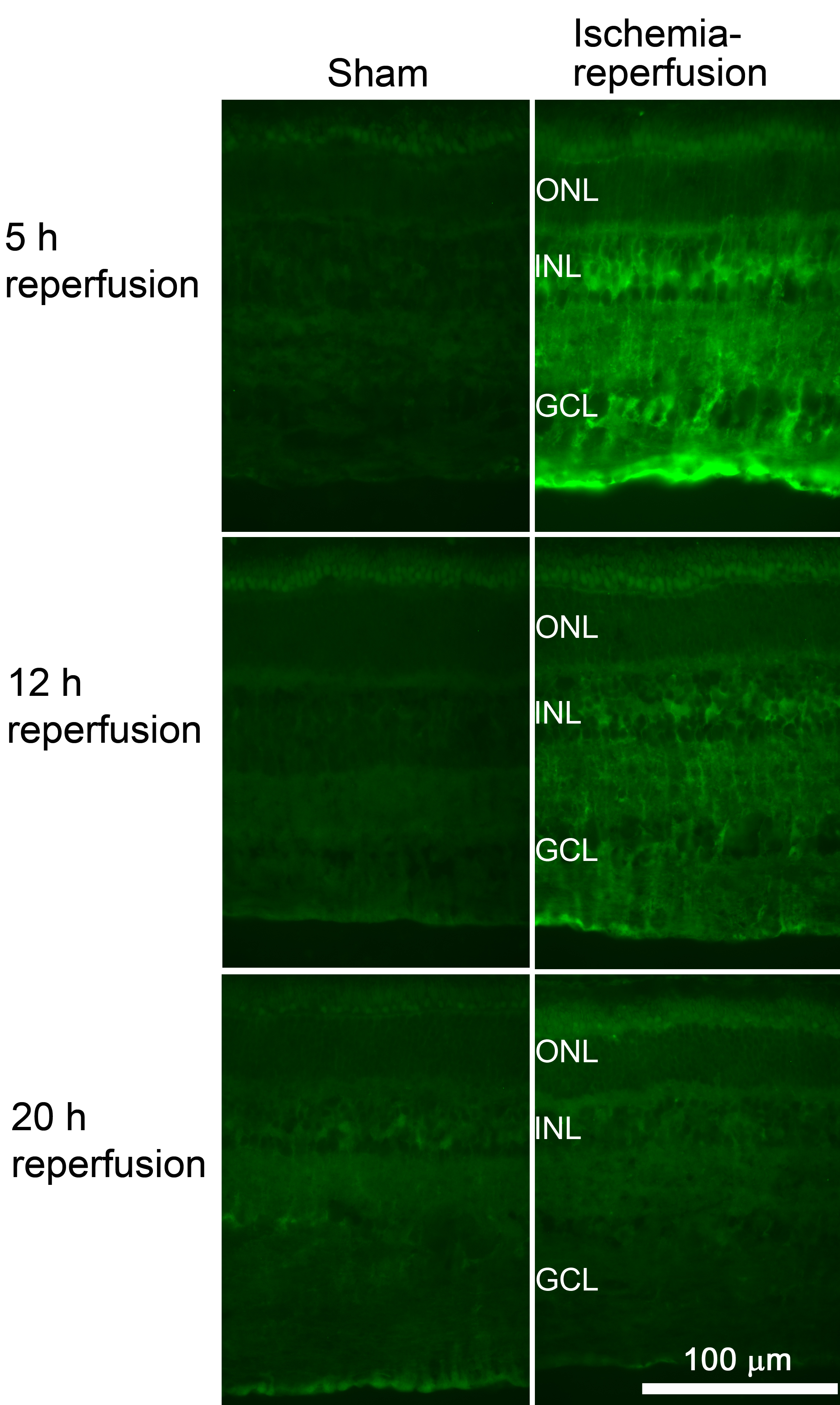
 Figure 4 of Gesslein, Mol Vis 2010; 16:392-407.