Figure 2. Terminal deoxynucleotidyl
transferase dUTP nick end labeling (TUNEL) staining in the neuroretina.
TUNEL staining of retinal sections from pigs subject to retinal
ischemia followed by 5, 12, or 20 h of reperfusion versus control
(sham). Note that TUNEL-positive cells can be detected in retinas from
ischemia-reperfusion eyes but are absent in retinas from sham-operated
eyes. The number of TUNEL-labeled cells increases gradually with the
duration of reperfusion. TUNEL-positive cells are observed throughout
the retinal sections, including the outer nuclear layer (ONL), inner
nuclear layer (INL), and ganglion cell layer (GCL). The bottom panels
are enlargements of the inserts above.
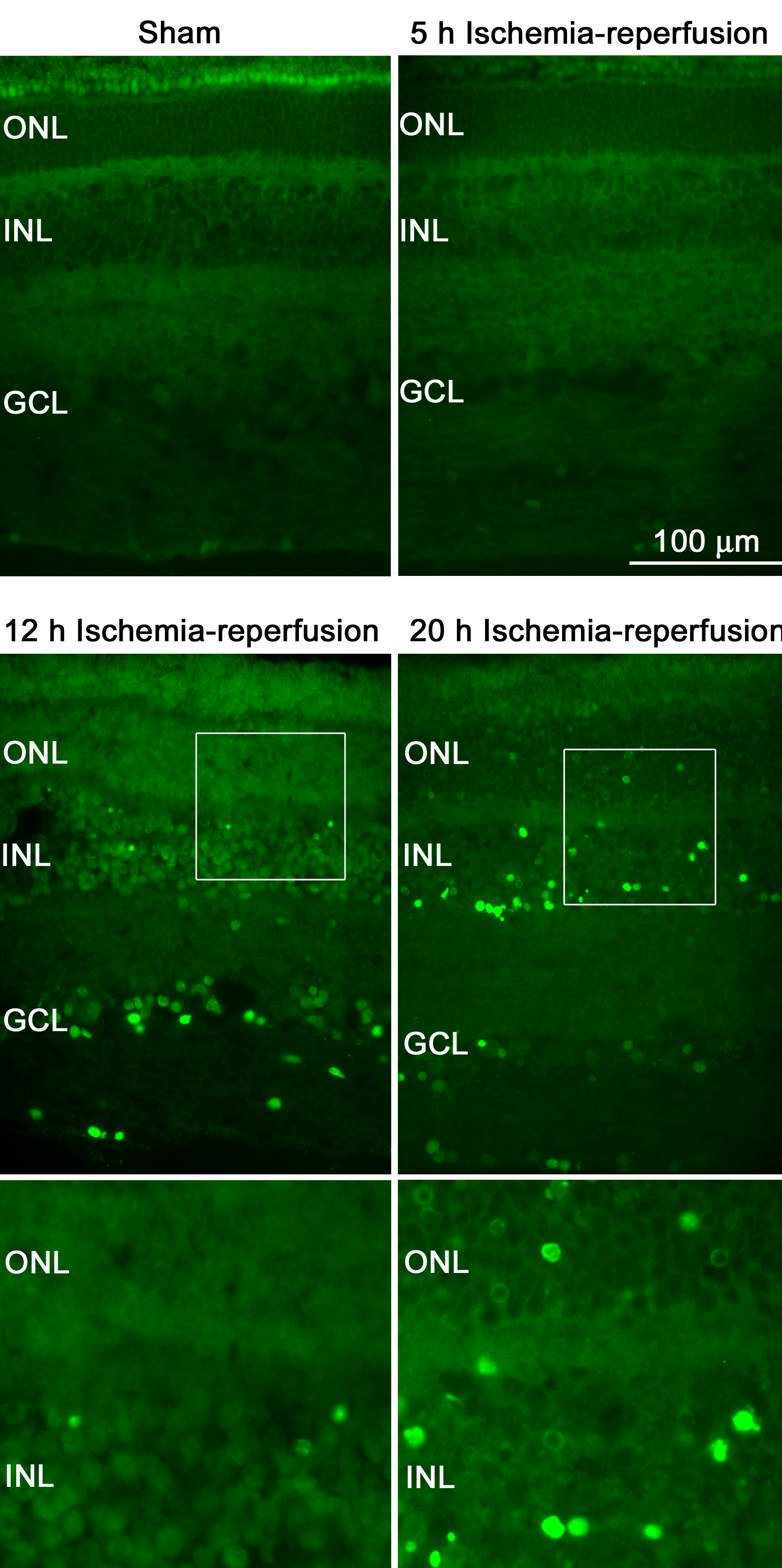
 Figure 2 of Gesslein, Mol Vis 2010; 16:392-407.