Figure 13. Total and phosphorylated c-jun
immunofluorescence in the retina. Representative examples showing
immunofluorescence staining of retina from pigs subjected to ischemia
followed by 5 (n=3), 12 (n=4), or 20 (n=3) h of reperfusion and the
corresponding sham-operated eyes. Note that the immunofluorescence
staining intensities for both total and phosphorylated c-jun are higher
in the ischemia-reperfusion eyes than in the sham-operated eyes. The
abbreviations used in the figure are outer nuclear layer (ONL), inner
nuclear layer (INL), and ganglion cell layer (GCL).
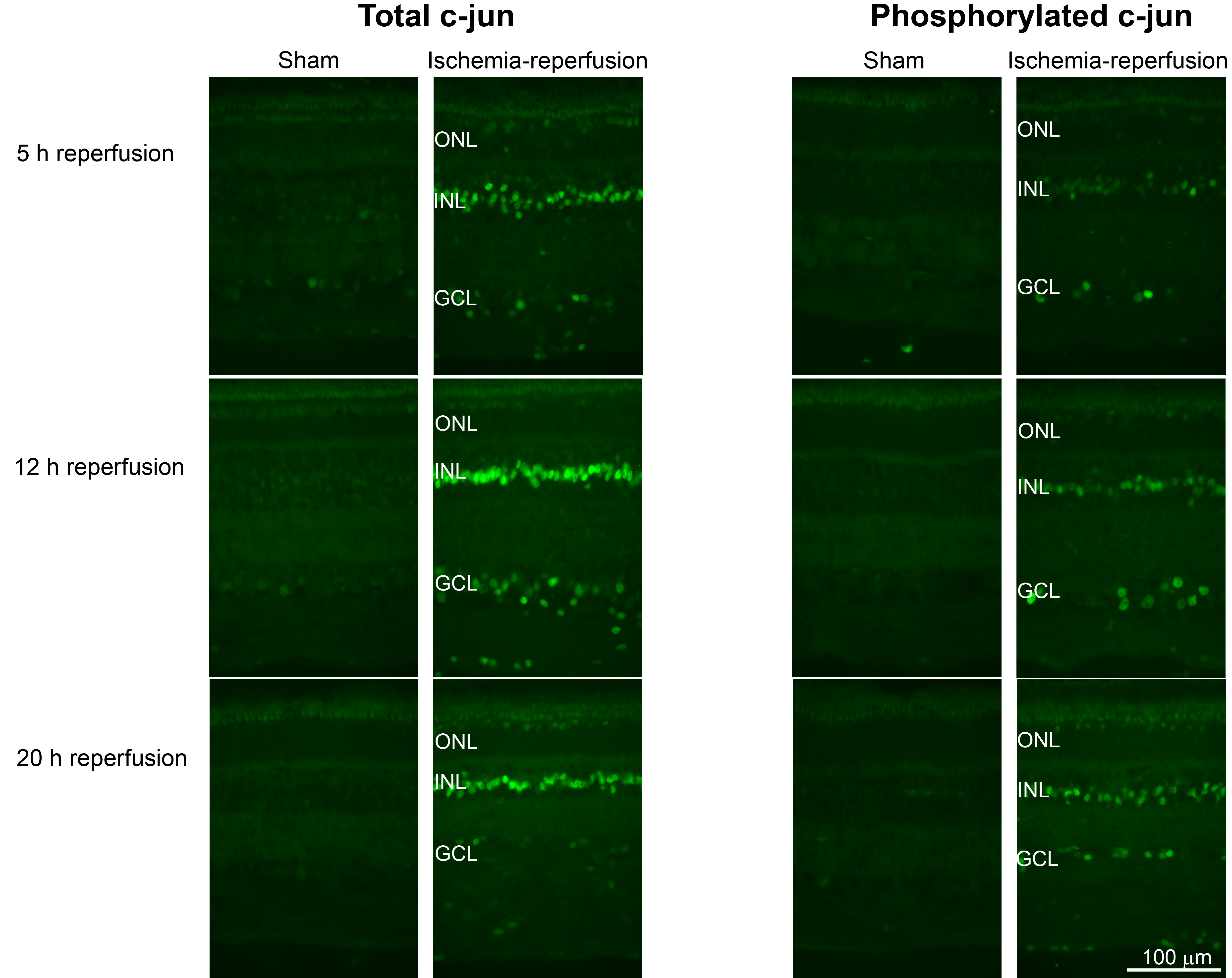
 Figure 13 of Gesslein, Mol Vis 2010; 16:392-407.