Figure 10. Phosphorylated
c-junNH2-terminal kinase 1, 2 and 3 (JNK1/2/3) immunofluorescence in
the retinal arteries and neuroretina. A: Representative example
of phosphorylated JNK staining of the retinal arteries. B:
Phosphorylated JNK was also detected in the ganglion cell layer (GCL)
and the inner nuclear layer (INL), but not in the outer nuclear layer
(ONL) of the neuroretina. Similar results were seen for sham-operated
and ischemia-reperfusion eyes.
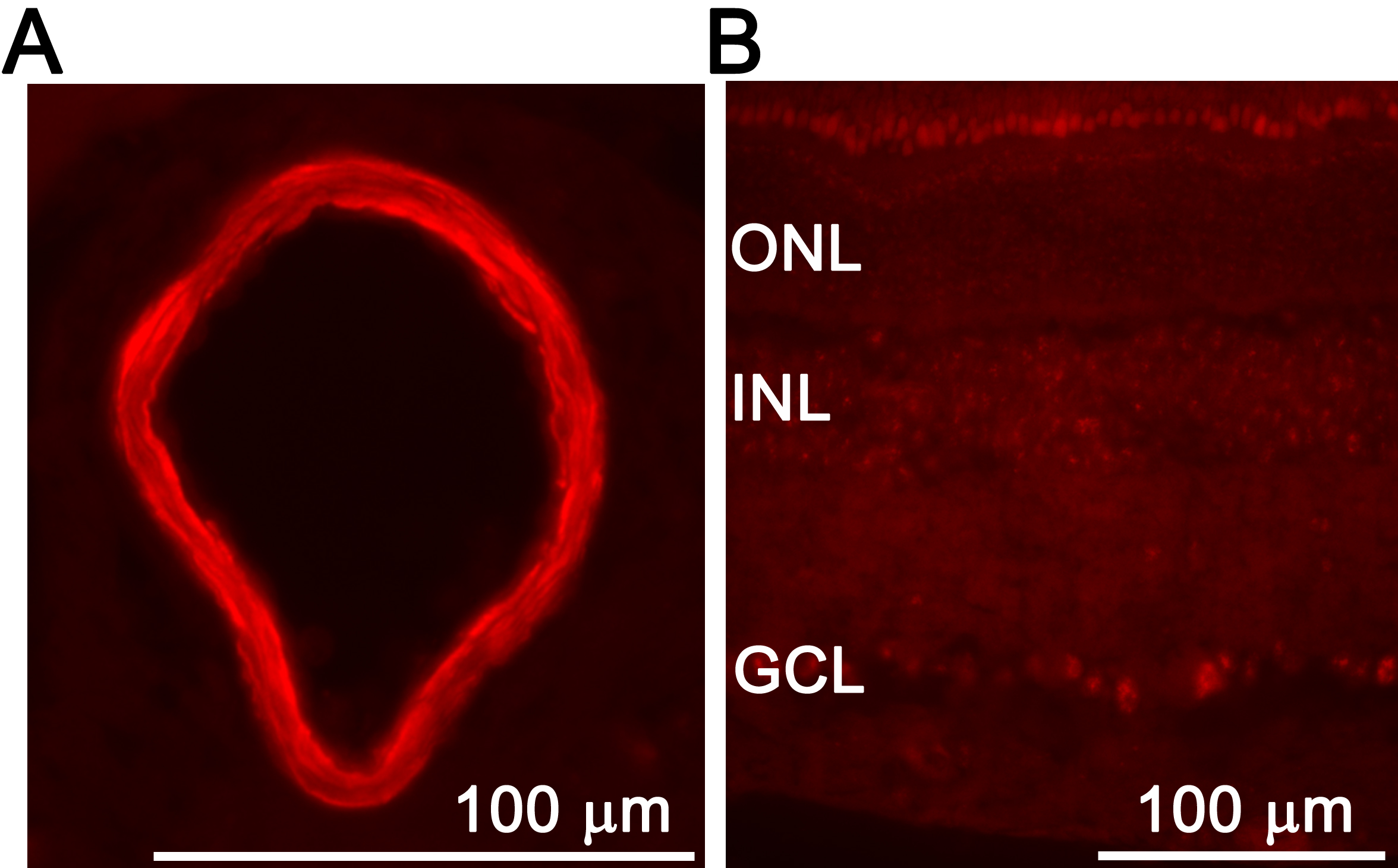
 Figure 10 of Gesslein, Mol Vis 2010; 16:392-407.