Figure 1. Pyknotic cells in the retinal
blood vessels and neuroretina. Histopathological examination was
performed by light microscopy of hematoxylin and eosin-stained sections
of porcine retina from the eye exposed to ischemia-reperfusion and the
sham-operated fellow eye. A: Artery and vein from an eye
exposed to ischemia-reperfusion and the corresponding sham-operated
eye. Pyknotic cell nuclei can occasionally be observed in the
ischemia-reperfusion eyes (arrows) but not in the sham-operated eyes. B:
Pyknotic
cell
nuclei
are observed throughout the retinal sections,
including the outer nuclear layer (ONL), inner nuclear layer (INL), and
ganglion cell layer (GCL) of the ischemia-reperfusion eyes (arrows) but
not in the corresponding sham-operated eyes.
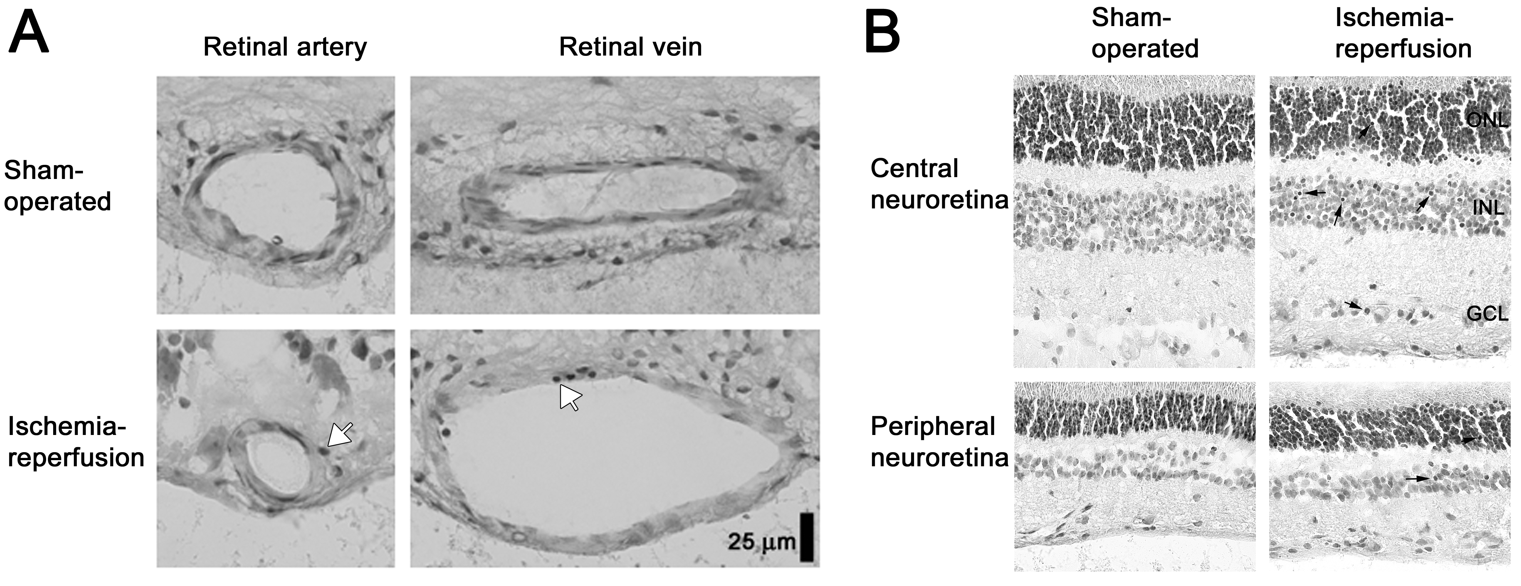
 Figure 1 of Gesslein, Mol Vis 2010; 16:392-407.