Figure 7. Single-flash electroretinograms of Rom1+/+, Rom1Rgsc1156/+, and Rom1Rgsc1156/Rgsc1156 mice. A: The amplitude of the a-wave differed significantly among the genotypes at every age examined (* indicates p<0.05, Tukey–Kramer
multiple-comparisons test). The reduction was most severe in the homozygotes. B: The amplitude of the b-wave also differed significantly among the genotypes (* indicates p<0.05, Tukey–Kramer multiple-comparisons
test), although there were no significant differences between the wild type and heterozygotes at 7 and 35 weeks. The number
of eyes analyzed in each group is indicated next to the symbols. Data shown are means±standard error.
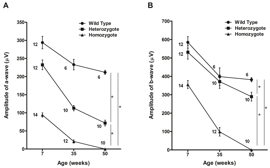
 Figure 7 of
Sato, Mol Vis 2010; 16:378-391.
Figure 7 of
Sato, Mol Vis 2010; 16:378-391.