Figure 6. Electron micrographs of retinas from 35-week-old Rom1+/+, Rom1Rgsc1156/+, and Rom1Rgsc1156/Rgsc1156 mice (n=3 each). The bar represents 10 µm in A, B, and C and 1 µm in D, E, and F. The photoreceptor outer segments were shorter in the heterozygotes (B) than in the wild-type mice (A). Remnants of the photoreceptor outer segments were observed in 35-week-old homozygotes (C, arrow). At high magnification the diameters of the discs clearly varied in the heterozygotes (E), and the discs of the heterozygotes were more compactly stacked than in the wild-type (D). (D-F) are high-magnified views of outer segment in (A-C), respectively.
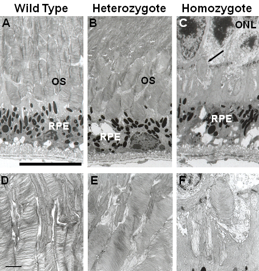
 Figure 6 of
Sato, Mol Vis 2010; 16:378-391.
Figure 6 of
Sato, Mol Vis 2010; 16:378-391.