Figure 4. Photomicrographs of retinas from 7- (A, B, C) and 35- (D, E, F) week-old Rom1+/+, Rom1Rgsc1156/+, and Rom1Rgsc1156/Rgsc1156 mice (n=3 or 4 each). The outer nuclear layer of the heterozygotes (B, E) was thinner and the photoreceptor outer segments were shorter than in wild-type mice (A, D). The alterations were even greater in the homozygotes (C, F). Differences were more marked at 35 weeks of age than at 7 weeks of age. GCL, IPL, INL, OPL, ONL, IS, OS, and RPE correspond
to ganglion cell layer, inner plexiform layer, inner nuclear layer, outer plexiform layer, outer nuclear layer, inner segment,
outer segment, and retinal pigment epithelium. The scale bar represents 50 µm.
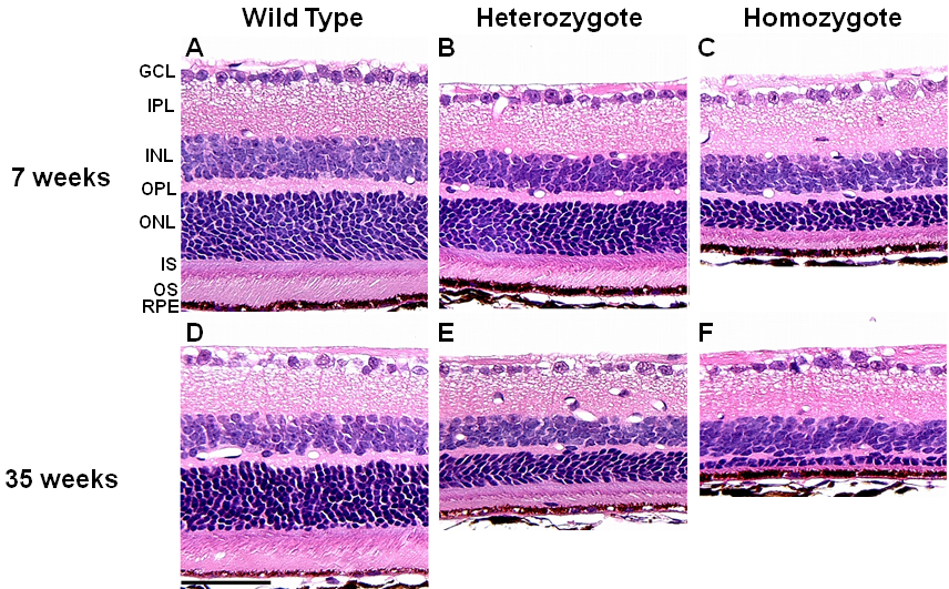
 Figure 4 of
Sato, Mol Vis 2010; 16:378-391.
Figure 4 of
Sato, Mol Vis 2010; 16:378-391.