Figure 11. Western blot analysis of retinal outer segment membrane protein 1 (Rom1), Prph2, and Rho in retinas from 3-week-old Rom1+/+, Rom1Rgsc1156/+, and Rom1Rgsc1156/Rgsc1156 mice (n=6 each). A: Western blots of 10 µg of retinal protein with antibodies to Rom1, Prph2, and β-actin. The stripped membrane was reprobed
with antibodies to Prph2 and β-actin. B: Western blots of 0.1 µg of retinal protein with antibodies to Rho and β-actin. The stripped membrane was reprobed with β-actin
antibodies. C: Semiquantitative analysis of Rom1, Prph2, and Rho levels. The western blot band intensities were measured with ImageJ software.
Rom1, Prph2, and Rho protein levels were normalized to β-actin levels. The intensity (mean±standard error) was normalized
to wild-type values, which were set equal to 100%. Protein levels differed significantly among the genotypes (* indicates
p<0.05, Tukey–Kramer multiple-comparisons test).
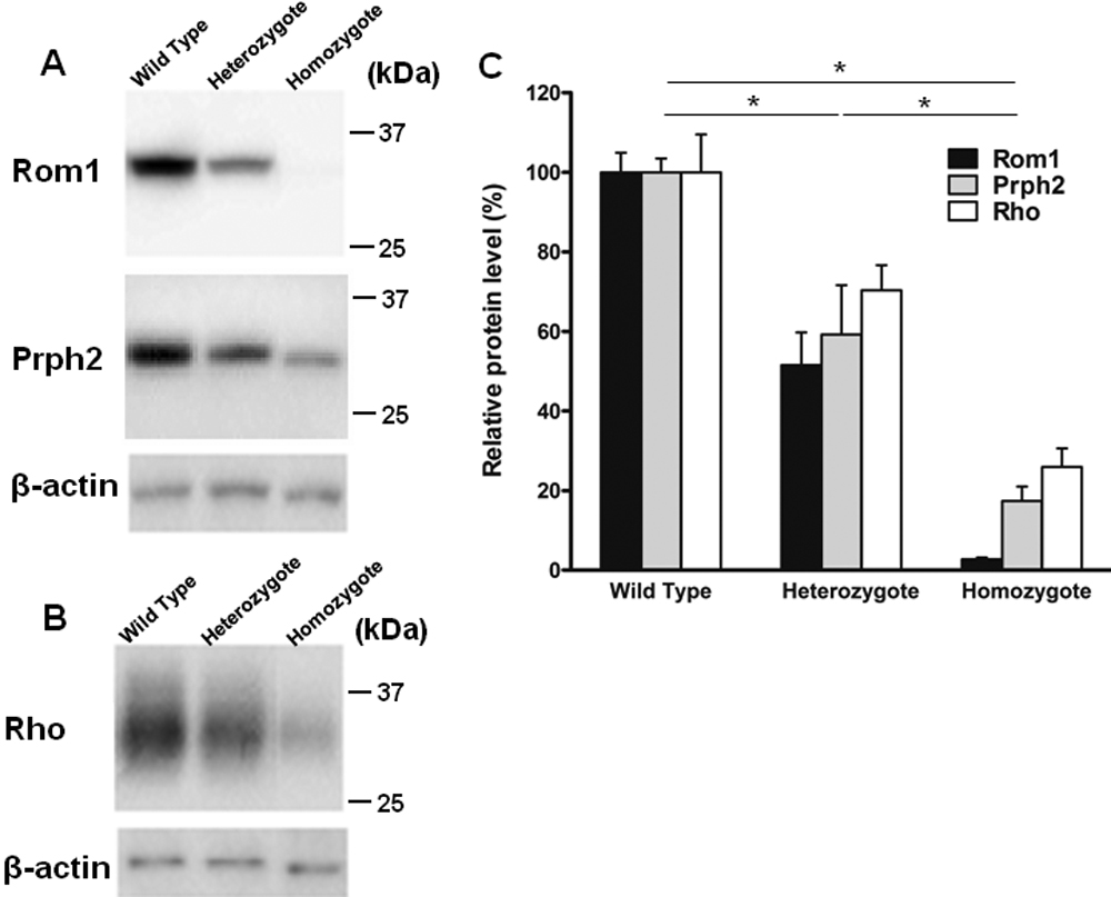
 Figure 11 of
Sato, Mol Vis 2010; 16:378-391.
Figure 11 of
Sato, Mol Vis 2010; 16:378-391.