Figure 1. Appearance of the fundus of an M-1156 mouse and a control mouse. At 45 weeks of age, the retinas of M-1156 mice were mottled and their arteries (indicated by arrows) were narrower than in the retinas of control mice which were F1
mice from crossing DBA/2J mice and C57BL/6J mice; DBF1. M-1156 mouse has same genetic background with DBF1.
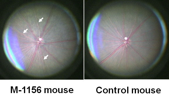
 Figure 1 of
Sato, Mol Vis 2010; 16:378-391.
Figure 1 of
Sato, Mol Vis 2010; 16:378-391.