Figure 1. Color fundus composite showing fundus autofluorescence (FAF) and spectral domain OCT (SD-OCT) for patients 1, 2, and 3. Patient
1 (Family A, Individual IV:2) at age 28: fundus image (A) shows well circumscribed macular atrophy, vascular attenuation, disc pallor, and peripheral pigment migration. FAF (B) using high gain demonstrates the absence of macular autofluorescence. SD-OCT (C) reveals thinning of the photoreceptor layer and wrinkling of the outer limiting membrane, with multiple high reflectance
bodies visible in residual outer nuclear layer. Patient 2 (Family A, Individual IV: 5) at age 12: fundus image (D) shows early macular atrophy and peripheral pigment migration. FAF (E) reveals limited parafoveal hyperfluorescence with low total autofluorescence. SD-OCT (F) reveals thinning of the photoreceptor layer, with discrete hyper-reflective bodies below the outer limiting membrane. Fundus
composite (G) of patient 3 at age 23 shows foveal and parafoveal yellow discoloration but minimal peripheral pigmentation. FAF (H) demonstrates hypofluorescence at the fovea and SD-OCT (I) shows thinning of the photoreceptor layer and high reflectance bodies.
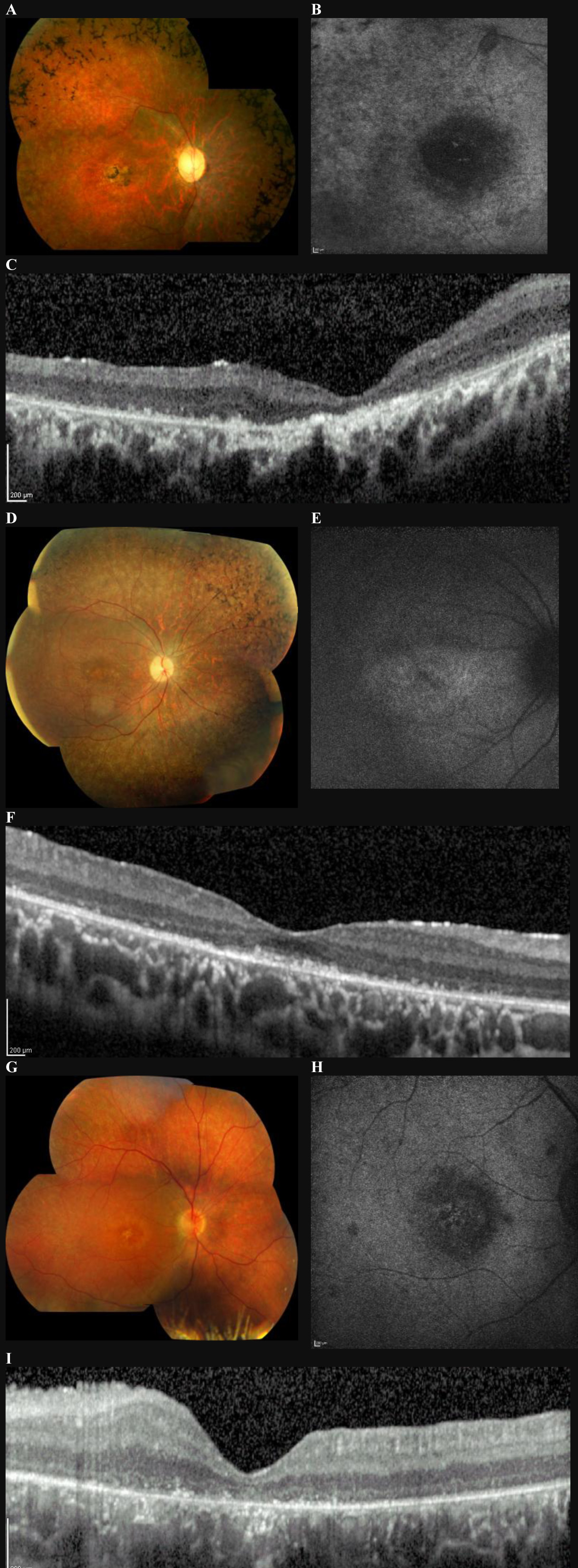
 Figure 1 of
Mackay, Mol Vis 2010; 16:369-377.
Figure 1 of
Mackay, Mol Vis 2010; 16:369-377.