Figure 2. Ultrastructural analysis of cells types in human corneal epithelium. A: Transmission electron micrograph (TEM) of terminally-differentiated corneal epithelial cells. Cells on the surface were
highly differentiated squamous cells with the most superficial being highly vesiculated with apoptotic nuclei and in the process
of desquamating (scale bar=1 μm). B: TEM of the transit-amplifying corneal epithelial cells. These basal cells are large columnar cells and contain large round
nuclei with diffuse chromatin (scale bar=1 μm). C: TEM of basal limbal stem cells. These cells are small with irregular nuclei that contain large amounts of condensed chromatin
(scale bar=1 μm).
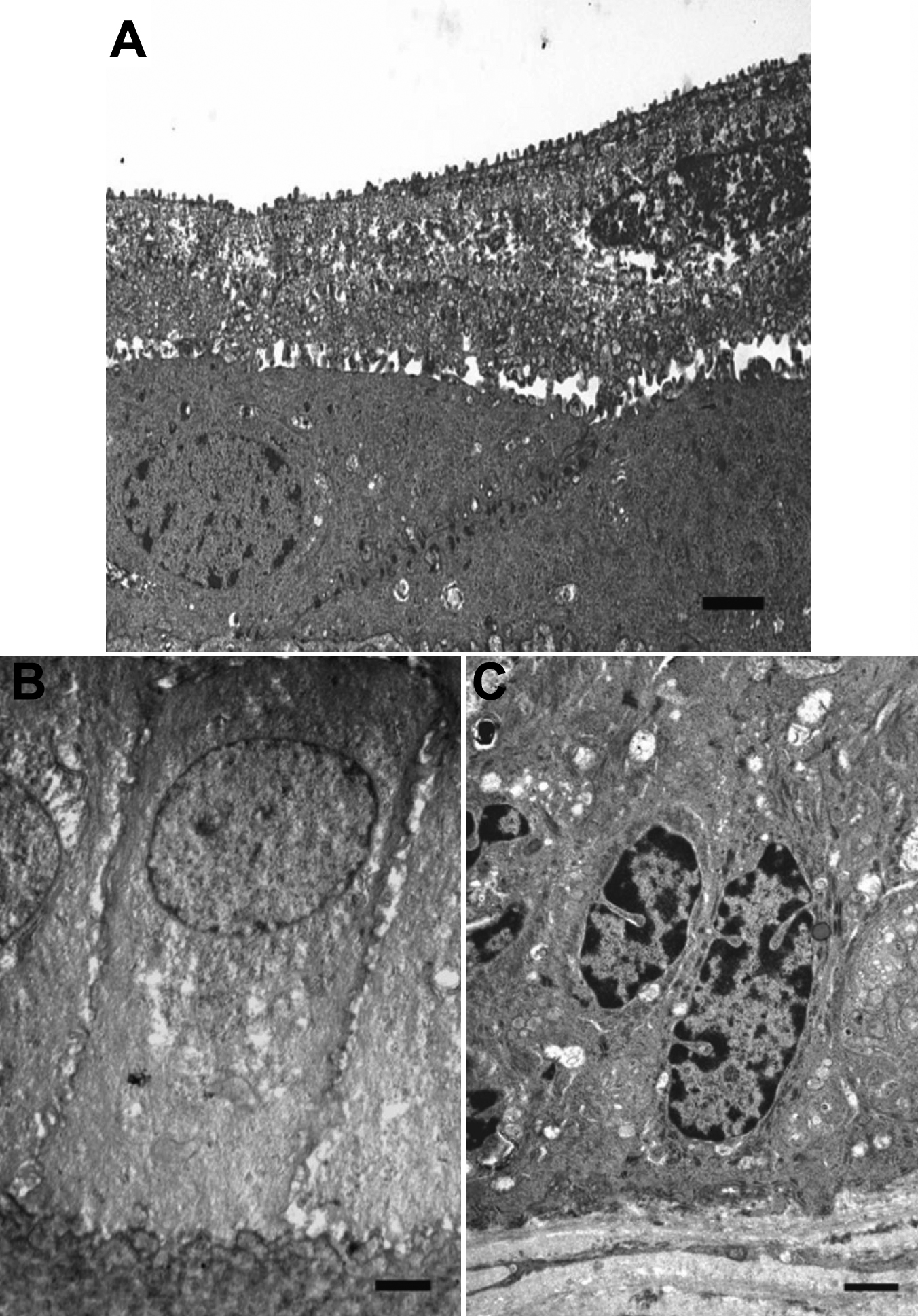
 Figure 2 of
Nakamura, Mol Vis 2010; 16:359-368.
Figure 2 of
Nakamura, Mol Vis 2010; 16:359-368.