Figure 7. The location of Foxe3 during the development of PCO in rat. Immunostaining for the Foxe3 protein (green) with nuclear counterstain
DAPI (blue). Foxe3 protein, normally confined to the anterior epithelial cells, is the same as in lens regeneration and as
it is in normal lens development. Note that the anterior capsule adheres to the posterior capsule on day 3 (A, B), no Foxe3 positive stain is found in the differentiating fibers at day7(C, D), and Foxe3 is strictly located in the lens epithelium on day14 (E, F) and 1 month after ECLE (G, H; I, J). Abbreviations: e, lens epithelium; f, lens fiber cells; t, transition zone.
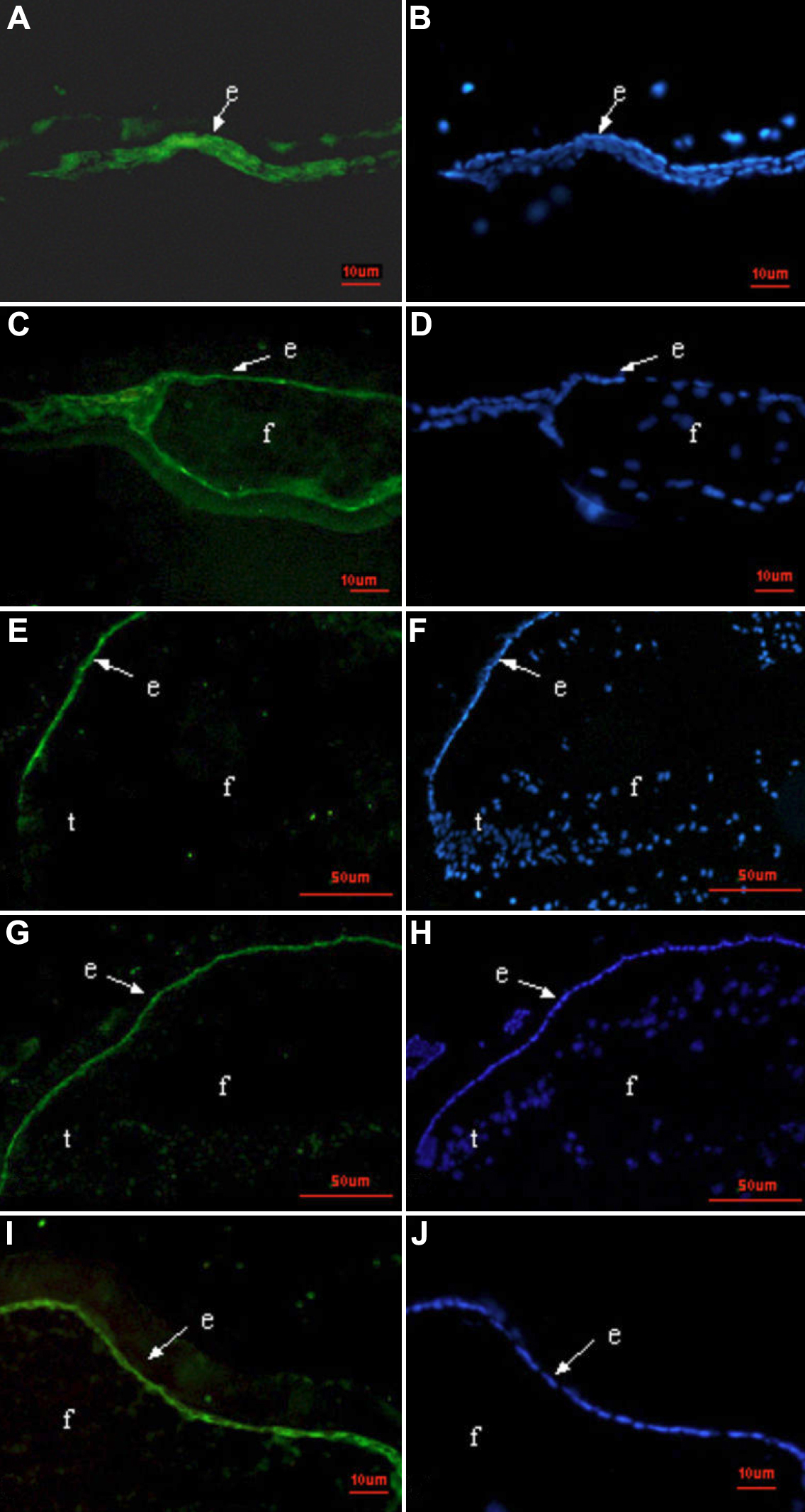
 Figure 7 of
Huang, Mol Vis 2010; 16:341-352.
Figure 7 of
Huang, Mol Vis 2010; 16:341-352.