Figure 6. Distribution of Prox1 protein
during the development of lens regeneration in a rat model.
Immunostaining for the Prox1 protein (green) with nuclear counterstain
DAPI (blue). Prox1 protein, normally found in epithelial cells and lens
fibers, appears similar in lens regeneration and normal lens
development. Note that the anterior capsule adheres to the posterior
capsule on day 3 (A, B), the fiber differentiation is
obvious on day 7 (C, D), and the Prox1 is found in the
LECs and differentiating lens fibers on day 14 (E, F).
One month after ECLE, Prox1 is mainly located in the nucleus of LECs
and to a lesser extent the cytoplasm of LECs in the region of anterior
capsule (G, H; I, J). While in the germinative
zone, Prox1 is absolutely located in the nucleus of LECs, in the lens
fiber cells, Prox1 is strictly located in the nucleus. Three months
after ECLE, Prox1 is mainly expressed in the transition zone (K,
L). Abbreviations: e, lens epithelium; f, lens fiber cells; t,
transition zone.
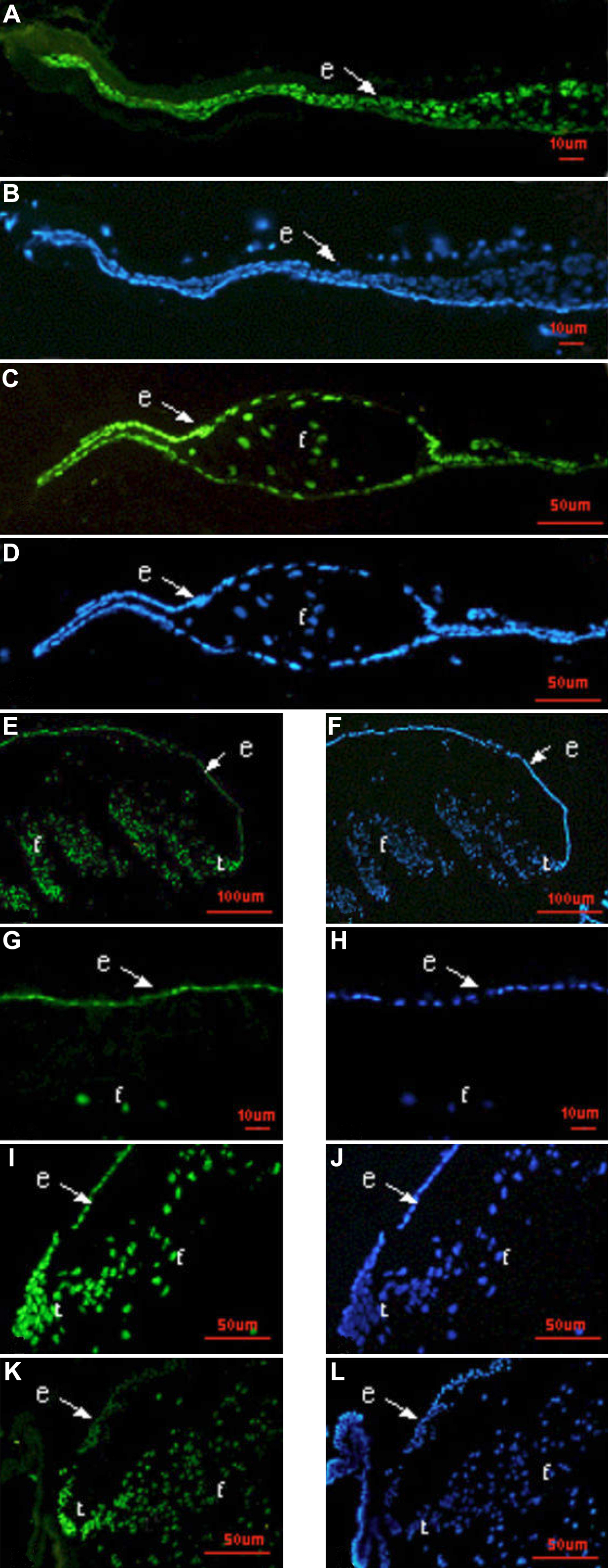
 Figure 6 of Huang, Mol Vis 2010; 16:341-352.