Figure 2. Hematoxylin and eosin staining
of lens tissue after ECLE. The capsular bag is open, and residual LECs
are observed on the peripheral anterior capsule immediately after ECLE (A).
On
day 3, LECs covered the inner surface of the anterior and posterior
capsules, and LECs lining the posterior capsule show early changes
characteristic of lens fiber differentiation (C). The LECs are
multilayered (D) and migrate to the central posterior capsule.
Some cells are spindle-shaped (E). On day 7, some cells are
spindle-shaped. The nuclei of LECs lining the posterior capsule
migrated away from the basement membrane (B).
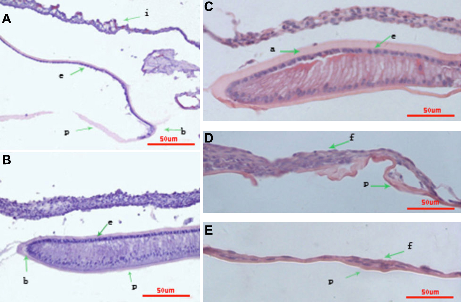
 Figure 2 of Huang, Mol Vis 2010; 16:341-352.