Figure 1. Morphological examination of rat
lens regeneration. Photographs of a rat eye (A, D, G,
J, M, P, S), lens tissue (B, E,
H, K, N, Q, T), and histologic analysis (C,
F, I, L, O, R, U;
hematoxylin and eosin staining) after ECLE. Continuous circular
capsulorhexis of the anterior capsule and clear posterior capsule in a
rat eye immediately after ECLE (A, B, C). On day
3, PCO is noted (D, E, F). On day 7, the eye
shows development of clinically evident PCO (G, H), and
Seomerring’s ring forms (G, H, I). On day 14,
elongated lens fibers on the posterior capsule become well
differentiated lens fibers (J, K, L).One month
after ECLE, the region of anterior capsulorhexis is opaque, and the
capsular bag is full of regenerated semitransparent lens material (M,
N, O). Two to three months after ECLE (P-U), the
regenerated lens is almost similar in size to the intact. Note the
relatively loose packing of the elongating fiber cells. Abbreviations:
a, anterior capsule; b, bow regions; c, cornea; e, lens epithelium; f,
lens fiber cells; i, iris; p, posterior capsule; r, retina.
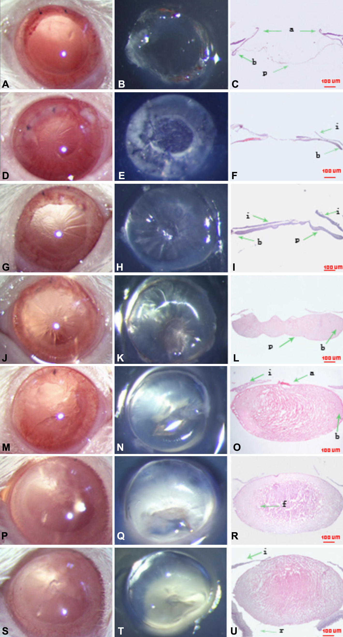
 Figure 1 of Huang, Mol Vis 2010; 16:341-352.