Figure 1. Effect of murine endostatin
(mEndo), murine-soluble vascular endothelial growth factor receptor 2
(msFlk-1), and murine-soluble Tie2 (msTie2) expression on proliferation
and migration of HUVEC cells. A: Equal numbers of HUVEC cells
were incubated for 24 h in viral supernatant from vehicle vector-PT67,
mEndo-PT67, msFlk-1-PT67, or msTie2-PT67 cells. The viable cell number
was measured by the conversion during 4 h of MTT into soluble formazan.
Bars represent standard deviation (SD; n=3 wells per measurement).
Similar results were obtained in three independent experiments. B:
HUVEC cells were seeded on 24-well tissue culture plates. When the
cells reached 90% confluence, a wound was made with a micropipette tip
in the center of the culture plates. The cultures were rinsed to remove
detached cells and incubated with medium containing viral supernatant
from vehicle vector-PT67, mEndo-PT67, msFlk-1-PT67, or msTie2-PT67
cells for 16 h. Digital images of wound closure were captured and used
for quantitative assessment of migration by measuring the distance
cells migrated beyond the injury lines. Four independent experiments
were conducted, and data were shown as mean±SD. The asterisk indicates
p<0.05, and the double asterisk indicates p<0.01, compared with
vehicle vector cells.
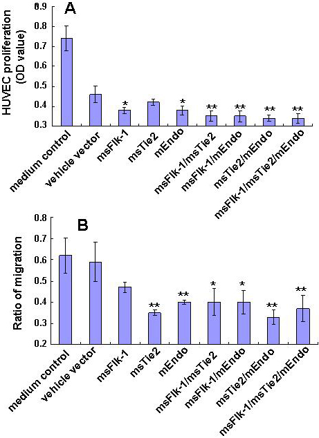
 Figure 1 of Chen, Mol Vis 2010; 16:310-319.