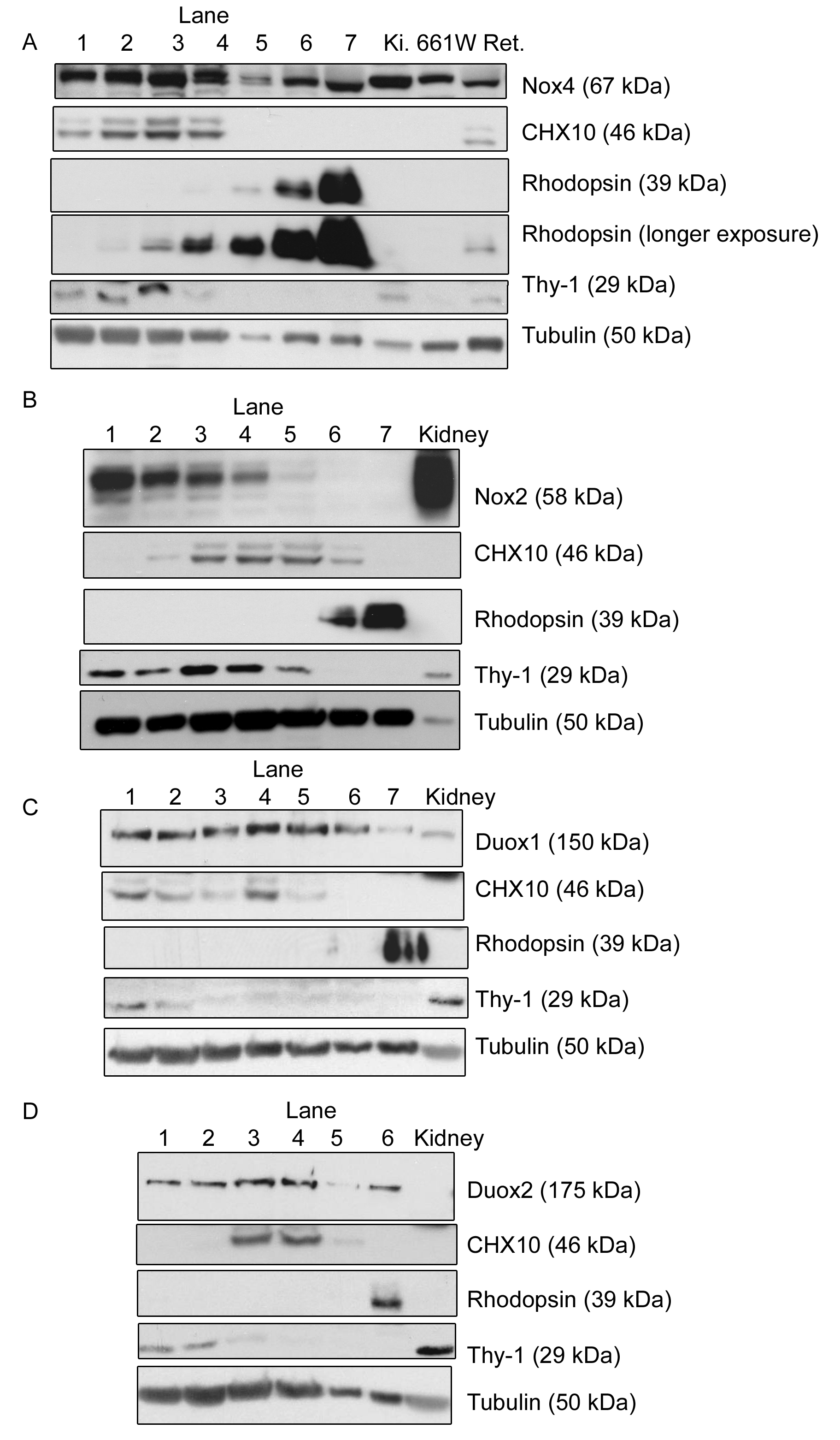
Figure 3. The expression pattern of
nicotinamide adenine dinucleotide phosphate oxidase (Nox) 4, Nox2, dual
oxidase (Duox) 1, and Duox2 in retinal layers is demonstrated through
western blotting of serial sections of the retina. In all parts, Lane 1
represents the first section removed from the ganglion cell side of the
retina, and lane 6/7 is the final section obtained from that retina and
represents the outermost layer of the photoreceptor cell side of the
retina. Thymocyte differentiation antigen 1 (Thy-1) served as a marker
of ganglion cells, rhodopsin of photoreceptor cells, and Ceh-10
homeodomain containing homolog (CHX10) of rod bipolar cells,
illustrating that the technique functioned well to separate the retinal
layers (middle panels of
A,
B,
C,
D).
In part
A, the three lanes on the far right of the gel are
control lanes, which are marked as follows, kidney (K
i.;
which is known to be a high expressor of Nox4), 661W cells, and a whole
retinal lysate (Ret.) to demonstrate the similarity between this and
our previous paper [
14].
In
B-
D, only the kidney control was maintained. The
expression of Nox4 throughout all of the retinal layers is demonstrated
(
A). Nox2 was expressed in most of the retinal layers, with
highest expression in the ganglion cell side and lowest at the
photoreceptor side (
B). Duox1 (
C) and Duox2 (
D)
showed similar expression patterns, with their highest expression
levels being in the middle lanes. Tubulin was used throughout as a
loading control. Blots are representative of at least three independent
experiments.
 Figure 3 of Bhatt, Mol Vis 2010; 16:283-293.