Figure 2. The reactive oxygen species (ROS) burst is generated in both rods and cones of the photoreceptor layer as determined by confocal
microscopy. Prior to imaging, retinal whole mounts were treated with either dihydroethidium (DHE)/peanut agglutinin (PNA;
A, B) or dihydrorhodamine (DHR)/PNA (C, D) for 60 min at 37 °C. Arrows point to the PNA-positive cones in red that produced ROS, as indicated by oxidized DHR123 staining
in green (D). These data were typical of at least three independent experiments. Confocal slices collected were 1.6 μm thick. The scale
bar represents 10 μm.
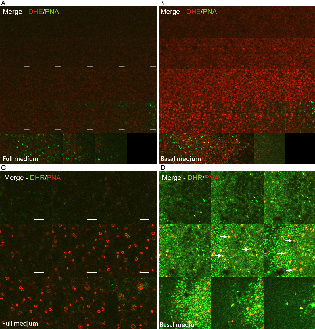
 Figure 2 of
Bhatt, Mol Vis 2010; 16:283-293.
Figure 2 of
Bhatt, Mol Vis 2010; 16:283-293.