Figure 1. Confocal microscopy in
conjunction with peanut agglutinin (PNA) staining allows
differentiation between rods and cones in live explants. A: A
schematic representation of the retina and its layers (ganglion cell
layer [GCL], inner plexiform layer [IPL], inner nuclear layer [INL],
outer plexiform layer [OPL], outer nuclear layer [ONL], inner and outer
segments [IOS]) to illustrate the difference between a transverse
section and a confocal slice. The image is a transverse section stained
with rhodamine-PNA and Hoechst to allow comparison with the confocal
images of B. The scale bar represents 25 μm. B:
Hoechst/PNA staining of the ONL on a whole-mount retinal explant by
confocal microscopy. Explants were cultured photoreceptor side facing
down. Retinal whole mounts were stained with Hoechst/PNA for 1 h at
37 °C before live imaging by an inverted confocal microscope. PNA,
the cone-specific marker, was used to bring the photoreceptor layer
into focus. Slices preceding the PNA-stained layers are the
Hoechst-positive nuclei in the ONL. These data are typical of at least
three different experiments. Confocal slices collected were 1.6 μm
thick. The scale bar represents 10 μm.
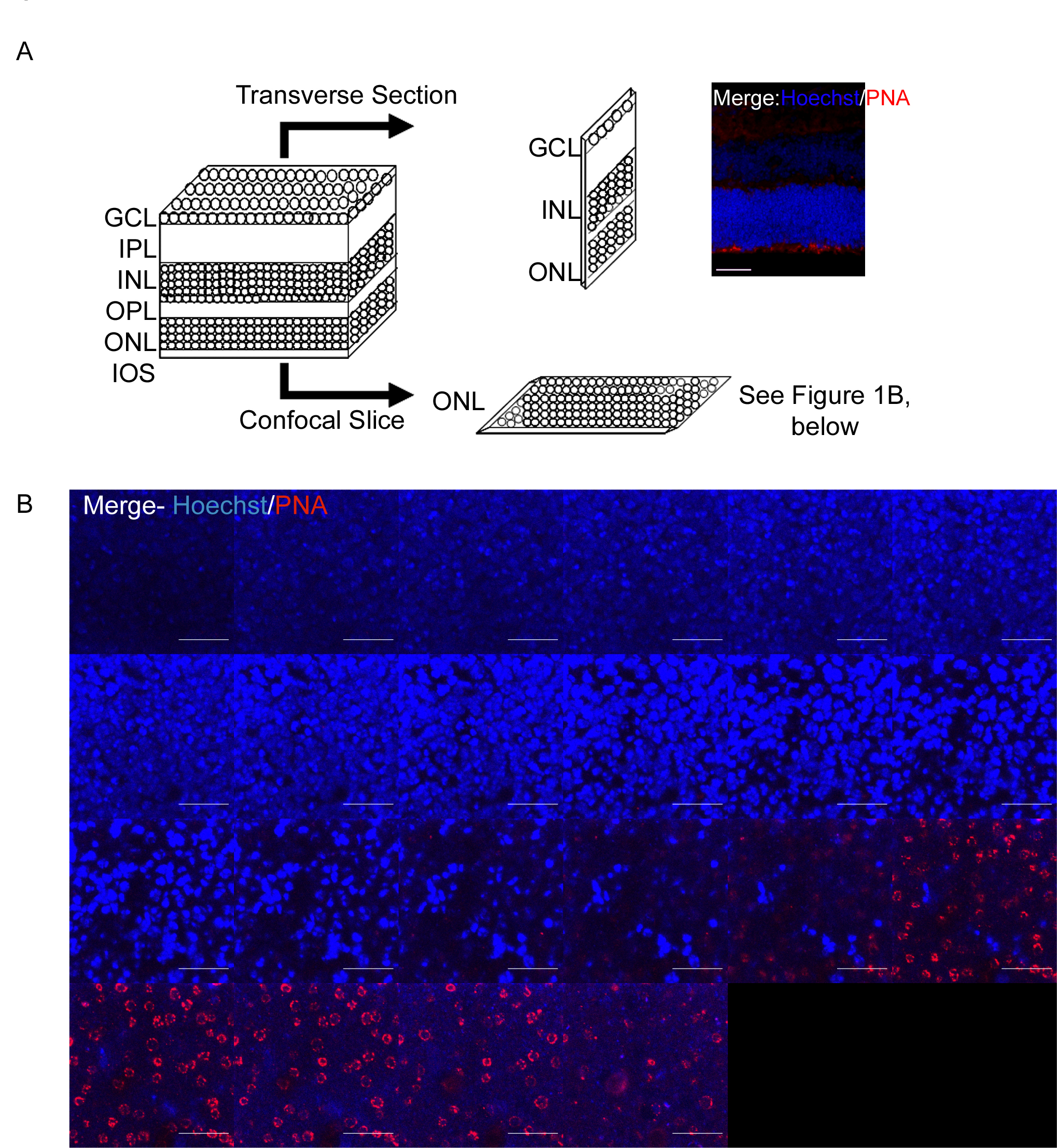
 Figure 1 of Bhatt, Mol Vis 2010; 16:283-293.