Figure 3. Family Q1 showing variable
phenotype. A: Pedigree of the family showing the splice site
mutation c.2240+1G>A and variable phenotypic presentation of the
affected members. Filled boxes represent affected individuals. Open
boxes represent unaffected individuals. Arrow indicates the proband. A
double line indicates presence of consanguinity in the family. B,
C: Representative slit lamp photomicrographs of the proband with
a homozygous splice site mutation c.2240+1G>A. The representative
clinical photographs of right (B) and left eye (C) of the
proband shows the presence of the typical ground glass appearance of
the cornea seen in autosomal recessive CHED. D: shows the
presence of apple green birefringence on staining with Congo-red and
viewing under polarized filter, marked by arrows. E: The slit
lamp photomicrograph of the right eye of the affected sibling had
marked stromal haze. F: The clinical photomicrograph of the
mother shows the endothelial deposits (marked by arrows) with stromal
haze. A few epithelial deposits are also seen.
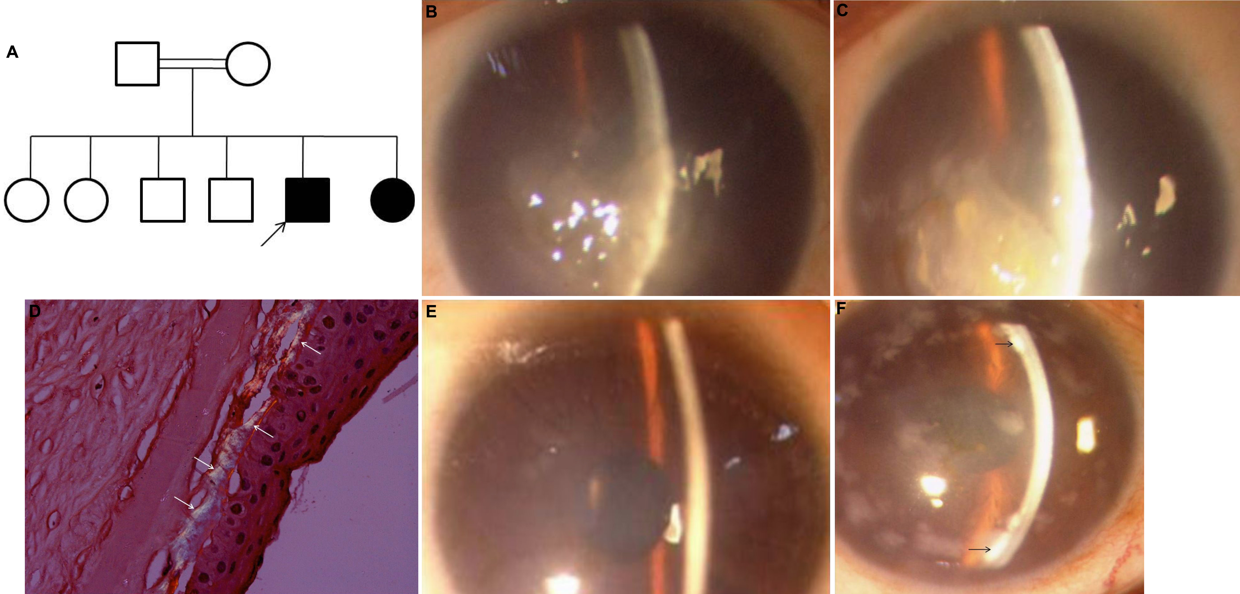
 Figure 3 of Paliwal, Mol Vis 2010; 16:2955-2963.