Figure 1. Segregation analysis of the
mutation c.672C>A (p.C224X) identified in family FRP-233. A:
Electropherogram corresponding to the wild type sequence (c.672C). B:
Electropherogram
corresponding
to the healthy mother, carrying the
mutation in heterozygous state (c.672C>A). C:
Electropherogram corresponding to the patient, carrying the mutation in
homozygous state (c.672A). Blue arrow indicates position c.672 is in A,
B and C, D: Family tree showing the segregation
of the p.C224X mutation.
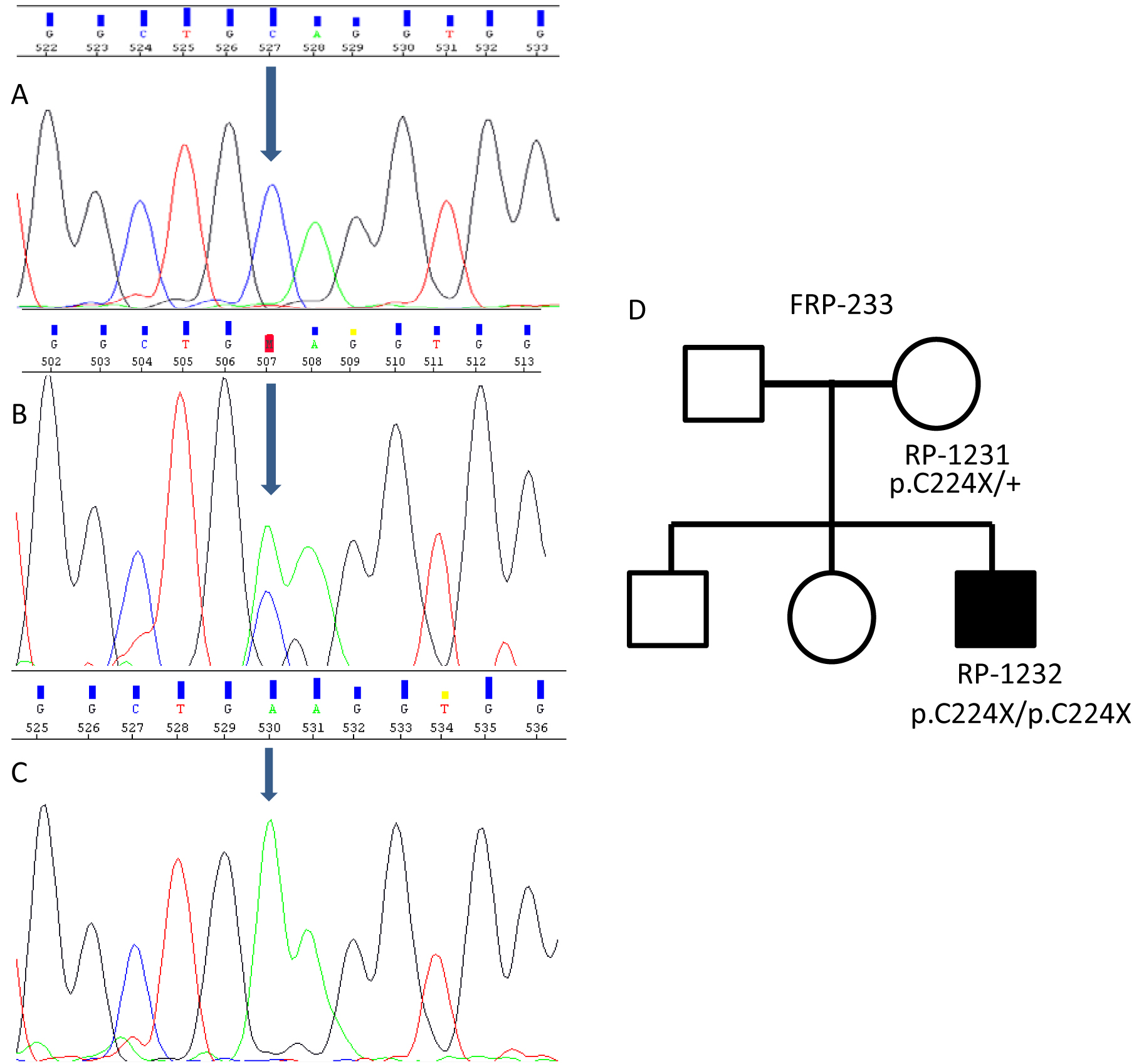
 Figure 1 of Aparisi, Mol Vis 2010; 16:2948-2954.