Figure 2. The ex vivo splicing assay. A:
Schematic
representation of the α-globin-fibronectin- extra domain B
(EDB) splice assay construct. Wild-type and mutant (c.102C>T) forms
of BEST1 exon 2 with flanking intronic sequence were cloned
into the α-globin-fibronectin-EDB splice assay vector. The position of
the mutated residue is highlighted with a star, and primer binding
sites to exonic vector sequences are indicated with arrows. B:
Splicing products generated by RT–PCR were separated by agarose gel
electrophoresis as indicated. The identity of the spliced products was
established by direct sequencing and is schematically represented on
the right. C: Agarose gel of RT–PCR reactions performed with
control primers designed against the vector sequence (dashed arrows in A)
demonstrates
equal loading of the cDNA template. The figure represents
results obtained from three separate experiments.
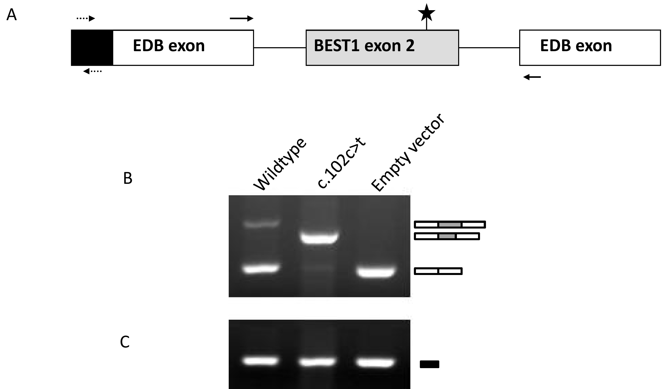
 Figure 2 of Davidson, Mol Vis 2010; 16:2916-2922.