Figure 1. Color fundus photographs showing
fundus autofluorescence (AF) imaging and horizontal spectral domain
optical coherence tomography (OCT) scans of affected individuals. A–C:
patient
1 at 44 years of age; D–F: patient 2 at 45 years
of age. Fundus pictures show widespread retinal pigment epithelium
(RPE) alterations and yellowish subretinal deposits along the vascular
arcades as well as yellow-white material in the maculae (A, D).
Changes
are more visible on AF imaging as diffuse, discrete areas of
hyper and hypoautofluorescence (B, E). On OCT,
intraretinal or subretinal fluids as well as atrophy are shown (C,
F).
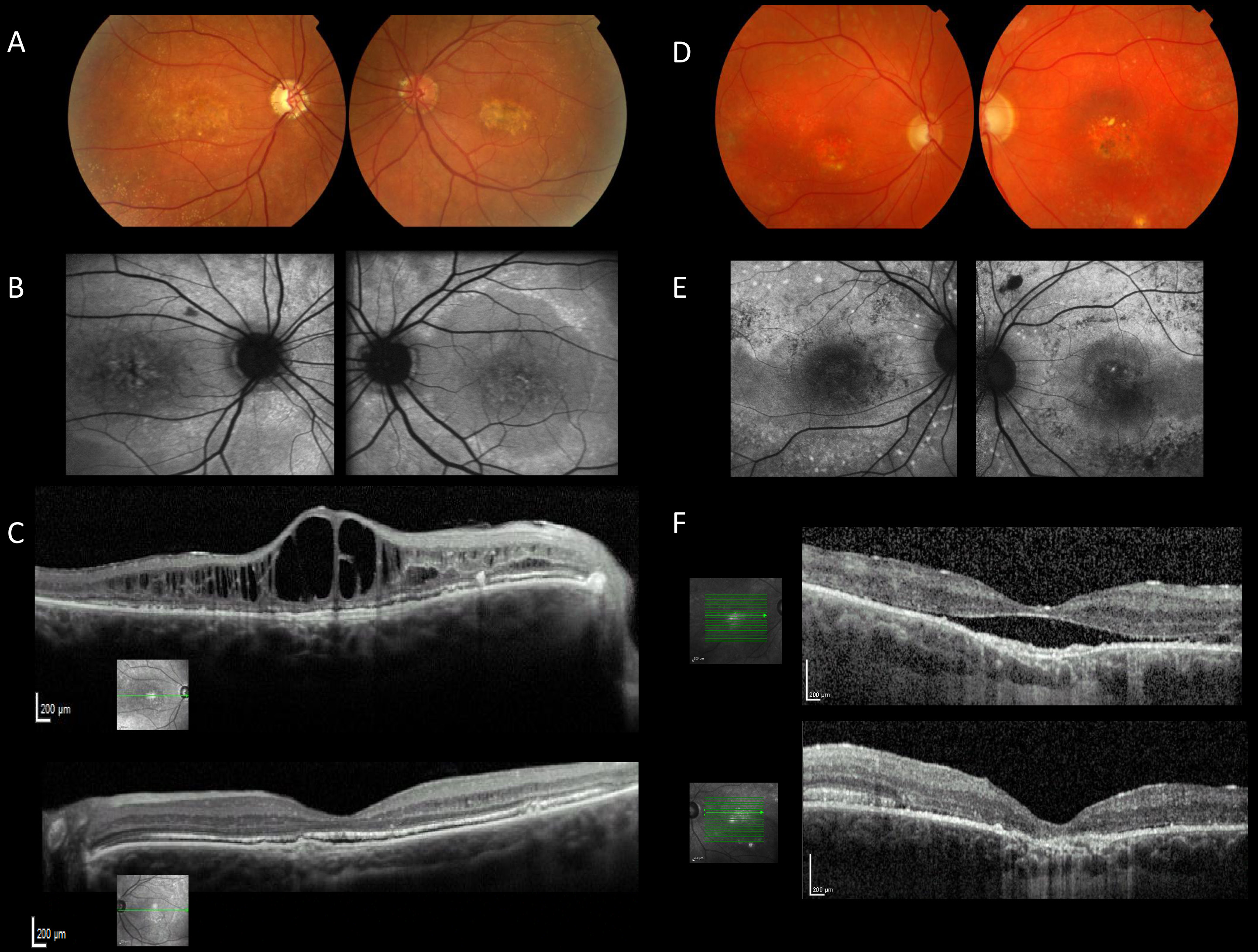
 Figure 1 of Davidson, Mol Vis 2010; 16:2916-2922.