Figure 5. The longitudinal sections of RGC axons were labeled with wheat germ agglutinin (WGA) for axonal regeneration. A: After optic nerve injury, the ends of axons became accumulated and twisted. B: One week after injured rich WGA-labeled axons were visualized at the proximal end of the traumatized area, a few axons entered
the distal end. C: One week after injury, the ends of the axons in the OEC+GDNF group were more extense and outspread. D: Two weeks after injury, some axons entered the traumatized area and reached the distal end of the traumatized area. E: Four weeks and F, 8 weeks after injury, WGA-labeled nerve fibers in the OEC+GDNF group were obviously prolongated and displayed abundant regenerated
axons. At these time points the figure showed regenerated axons tangled and fluorescence accumulated there. G: Eight weeks after injury, a few new fibers regenerated in the OEC group and extended across the traumatized area to reach
the distal end; however, the regenerated axons appeared significantly less than in the OEC+GDNF group. H: Negative control for WGA-labeled optic nerve axons. All images: left is the proximal end and right is distal end of optic
nerve injured point. The scale bar is 100 μm.
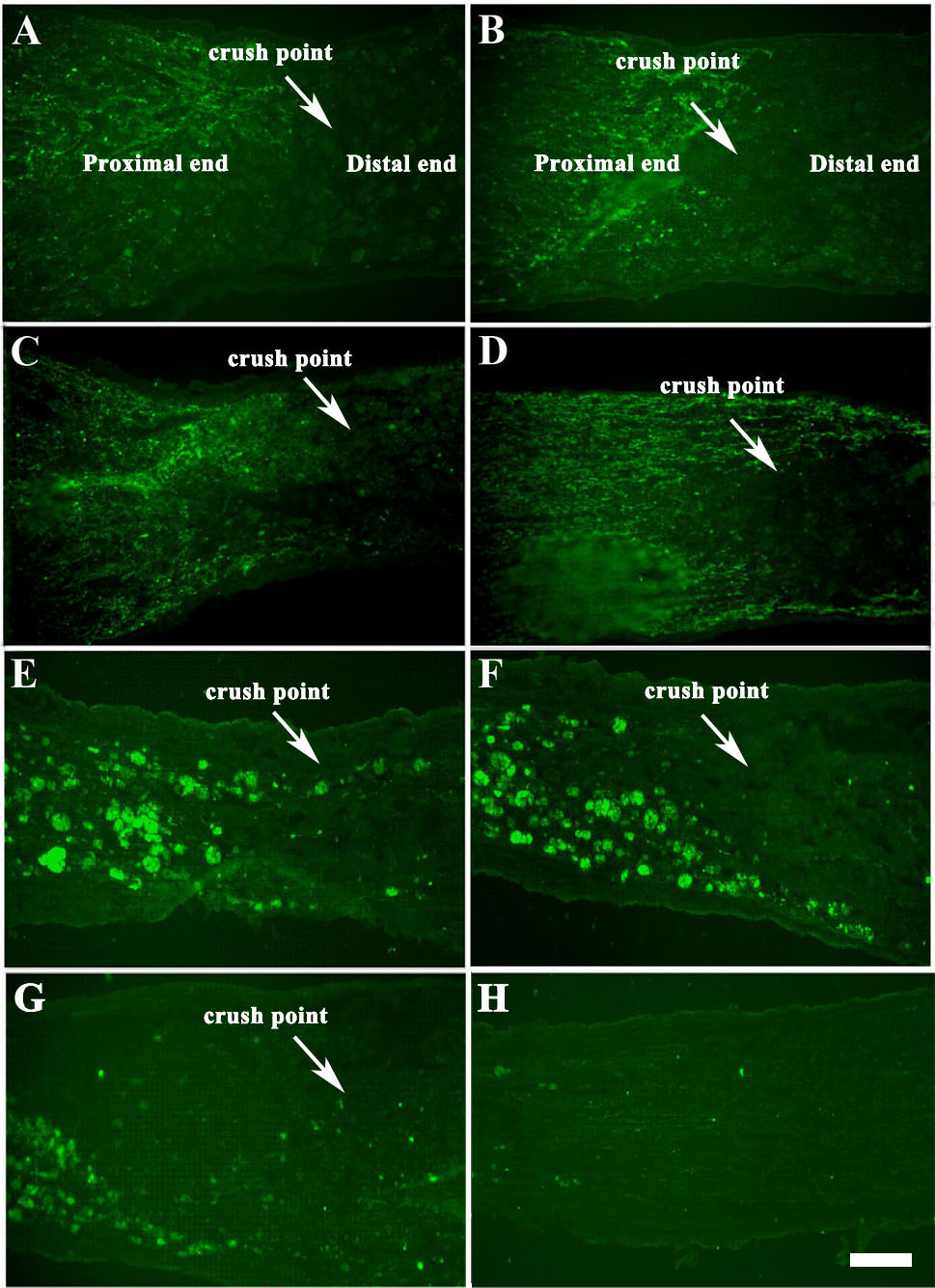
 Figure 5 of
Liu, Mol Vis 2010; 16:2903-2910.
Figure 5 of
Liu, Mol Vis 2010; 16:2903-2910.