Figure 2. Space-filling model in four
orientations for modified myocilin proteins. Full length myocilin,
deletions due to presence of stop codon and deletions/modifications due
to possible alternative splicing caused by the single nucleotide
polymorphism shown in the model, as explained in Figure 1. Different
regions in the model are colored as follows; NH2-terminal
region (Orange), coiled coil region (Pink), hinge region (Cyan),
COOH-terminal region (Yellow), regions predicted to be deleted (White)
due to stop codon mutation or possible alternative splicing, regions
predicted to be modified due to possible alternative splicing.
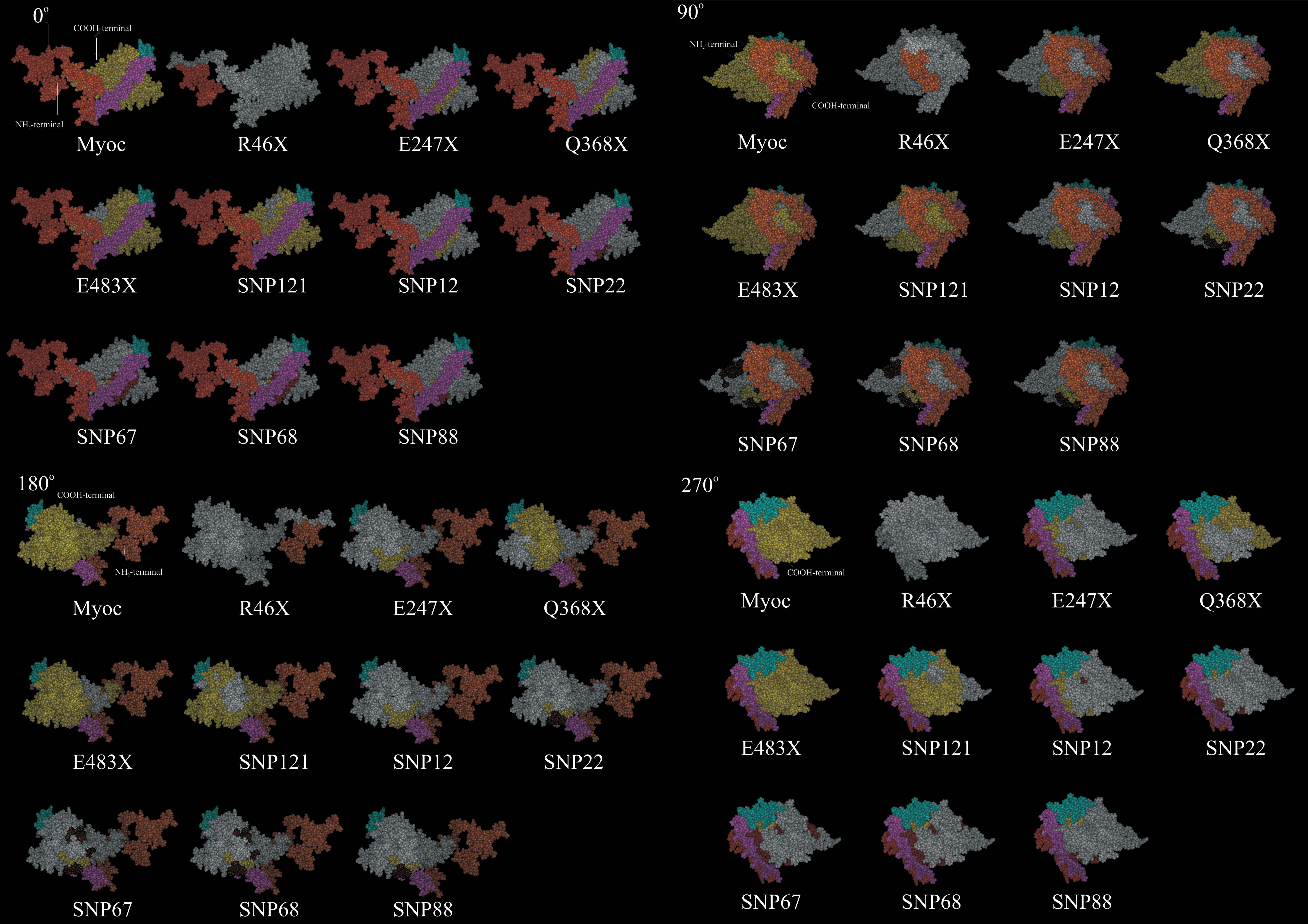
 Figure 2 of Pandaranayaka, Mol Vis 2010; 16:2891-2902.