Figure 5. Treatment by ATP-liposomes
results in neuroprotective effects in the ganglion cell layer of
retinas after ischemia reperfusion. A: The percentage survival
of ganglion cell layer (GCL) neurons in the ischemic retinas of ATP
(ATP)-, phosphatidylcholine (PC)-liposome-, and PBS-treated animals
were detected 7 days after reperfusion (*p<0.05, **p<0.01, n=6). B:
The
percentage of Neuronal Nuclei (NeuN)-labeled neurons in regions of
central, middle, and peripheral retina were compared between
sham-operated (sham) and ischemic eyes of ATP-, PC liposome-, and
PBS-treated animals 7 days after reperfusion (*p<0.05, **p<0.01,
n=6). C: Representative confocal images of NeuN-labeled GCLs
(green) in flatmounted controls (sham) and ischemic retinas were taken
7 days after reperfusion. Scale bar indicates 100 μm.
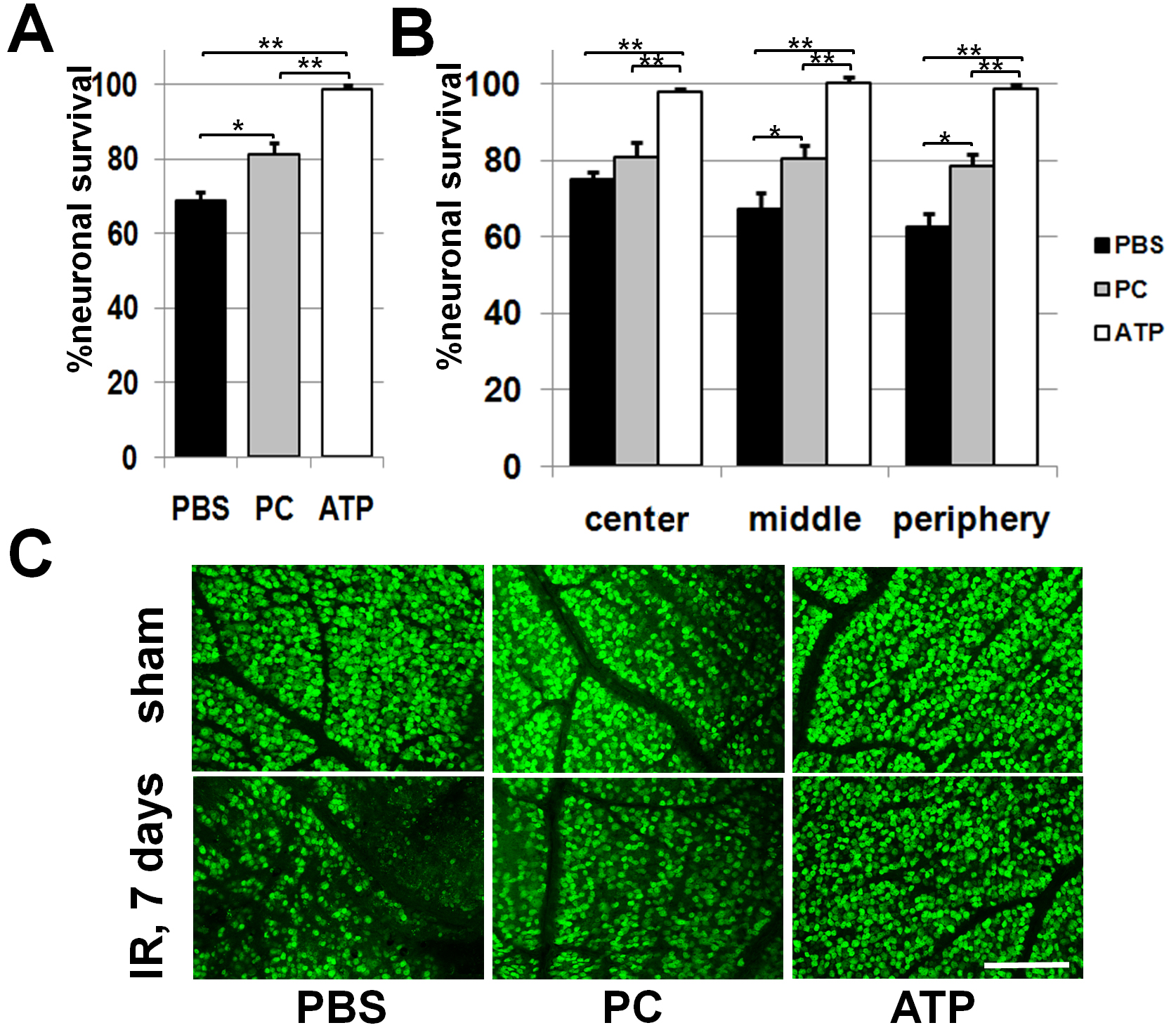
 Figure 5 of Dvoriantchikova, Mol Vis 2010; 16:2882-2890.