Figure 3. ATP-liposomes prevent retinal
ischemia-induced necrosis. A: Transmission electron microscopic
(TEM) analysis of the ganglion cell layer (GCL) of sham operated (sham)
and ischemic retinas treated with ATP (ATP)-, phosphatidylcholine
(PC)-liposomes, and PBS 24 h after reperfusion was used to identify
necrotic and apoptotic cells. B: Quantitative comparisons in
terms of the cell populations showed characteristic features of
necrotic (closed column) and apoptotic (open column) cells detected by
the analysis of the TEM micrographs. Results represent the
mean±standard error of the mean of three to five independent
experiments (*p<0.05).
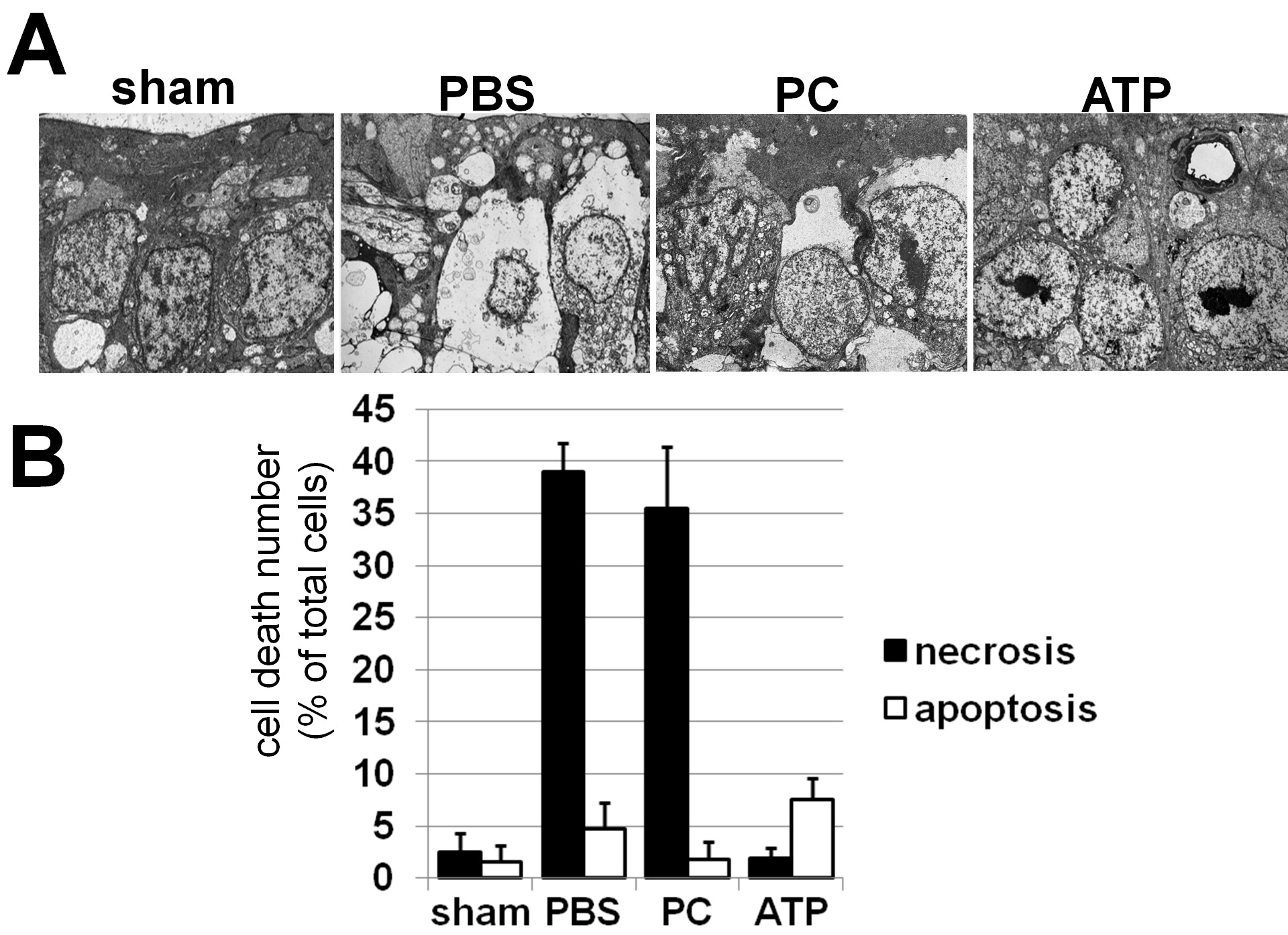
 Figure 3 of Dvoriantchikova, Mol Vis 2010; 16:2882-2890.