Figure 2. ATP-liposomes effectively pass
through the blood–retinal barrier. A: Confocal photomicrographs
of flatmounted ischemic retinas treated with carboxyfluorescein
(CF)-liposomes or carrier buffer PBS were taken 24 h after ischemia
reperfusion (IR). Nonfixed freshly isolated retinas were visualized by
confocal microscopy in the ganglion cell layer (GCL). Retinal GCL cells
display green fluorescence, indicating liposomal uptake (yellow
arrows). Punctuate traces of CF labeling are likely endothelial cells
in blood capillaries (red arrows). PBS-treated controls display little
autofluorescence. The scale bar represents 50 µm. B: Relative
changes in ATP (ATP) content in control (sham) and ischemic (24 h)
retinas following the treatment with ATP, PC- liposomes, and PBS 24 h
after reperfusion. The results are shown as a percentage of the
corresponding value in PBS-treated sham-operated retinas (*p<0.05,
n=5).
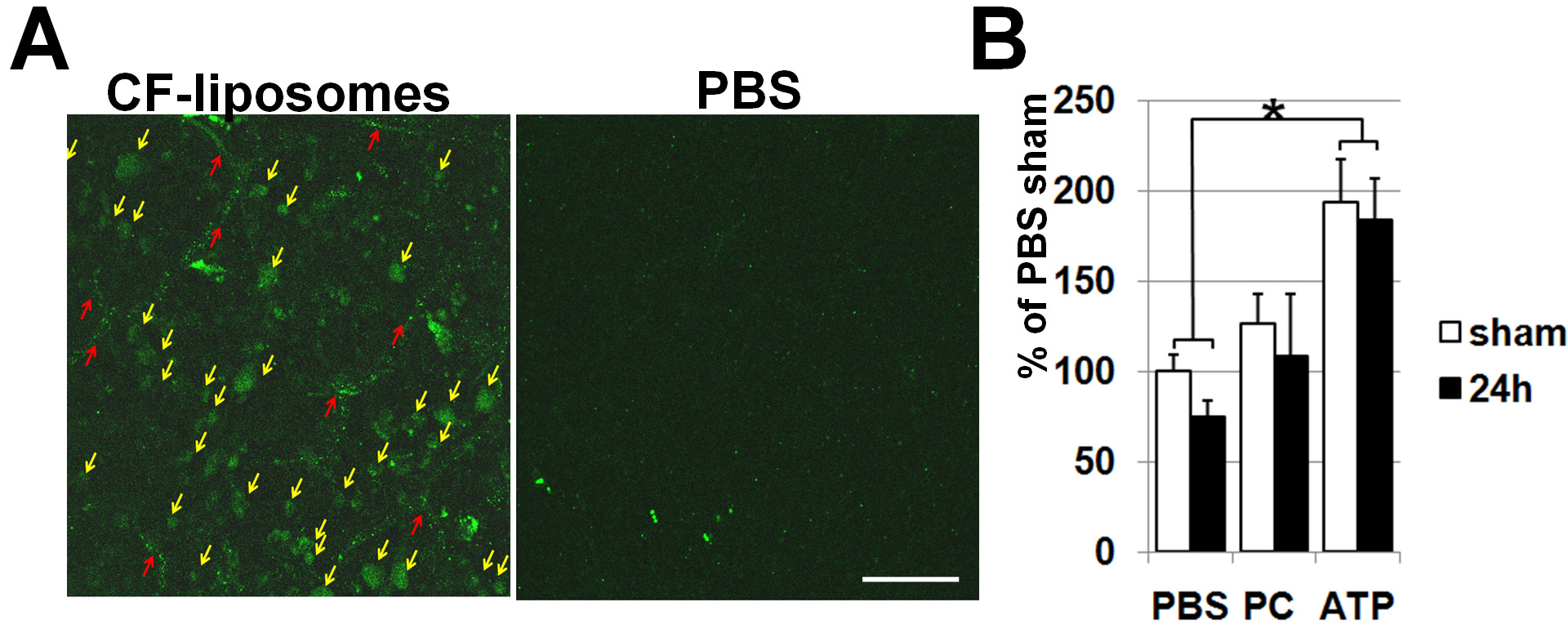
 Figure 2 of Dvoriantchikova, Mol Vis 2010; 16:2882-2890.