Figure 4. Expression of MerTK in retinal
pigmented epithelium (RPE) of wild-type and Nrl−/−
mice. In wild-type mice, the photoreceptor outer segment (POS) is
intensely stained by rhodopsin antibody (A), and MerTK overlaps
to a large extent with this stain (B). However, the MerTK label
does not extend all the way to the base of the photoreceptor OS, so the
merged image (C) shows a mixture of green (overlapping stain)
and green (rhodopsin only). This is further illustrated by the two
arrows (white to show the width of the rhodopsin stain, gray to show
the width of the MerTK stain). S-cone opsin immunostaining in Nrl−/−
retinas shows aligned short POS (D), and the pattern of
MerTK expression in Nrl−/− RPE is much reduced,
compared to wild-type retinas, appearing as a discontinuous thin band
running along the interface between the RPE and outer nuclear layer
(ONL; arrow in E). F: The general structure of the Nrl−/−
retina is seen by 4,6-di-amino-phenyl-indolamine (DAPI) staining of
nuclei, showing the monolayer of rounded nuclei within the RPE (arrow).
Scale bar represents 10 µm.
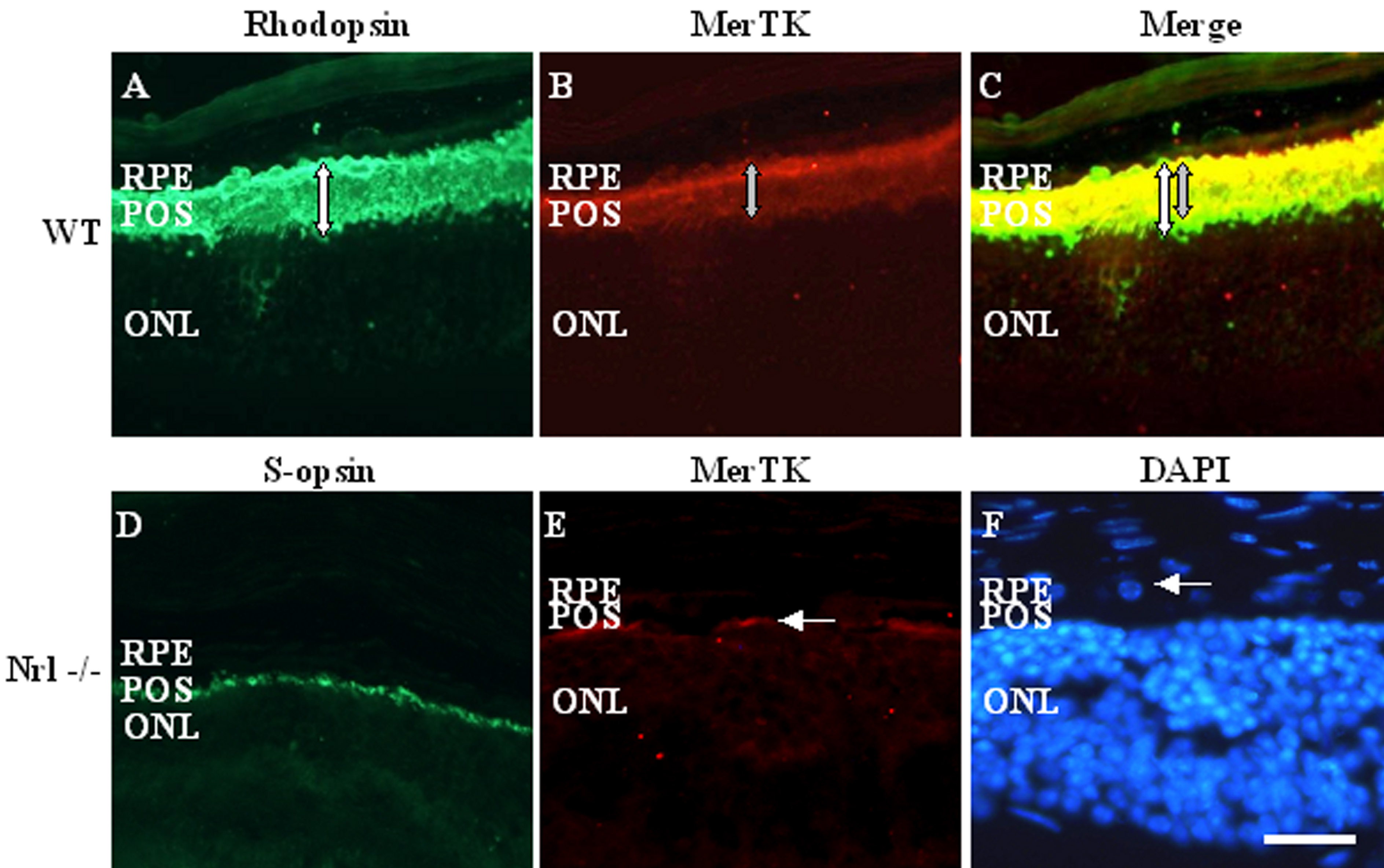
 Figure 4 of Krigel, Mol Vis 2010; 16:2873-2881.