Figure 2. Immunohistochemical detection of
rhythmic phagocytosis in short wavelength sensitive (S)-cones of young
adult Nrl−/− and wild-type mice. A and B:
These
two sections were taken from animals killed under light-dark
conditions at, respectively, hour 1 and hour 10 of zeitgeber time. C
and D: These two sections were taken from animals killed under
total darkness conditions at, respectively, hour 1 and hour 10
circadian time.. Sections immunolabeled with S-cone opsin antibody show
the continuous row of cone outer segments across the section, and the
presence of numerous immunoreactive inclusions (phagosomes=Ph) within
the retinal pigment epithelium (RPE; short arrows) at ZT1 and
CT1, but not at ZT10 and CT10. E: S-cone opsin immunostaining
of wild-type mouse retina (ZT6) shows the presence of occasional
labeled COS (long arrows). RPE, retinal pigment epithelium (the
approximate level of the basal surface of the RPE) is shown by a dashed
line in panels A-D. Scale bar in E represents 10
μm for all panels.
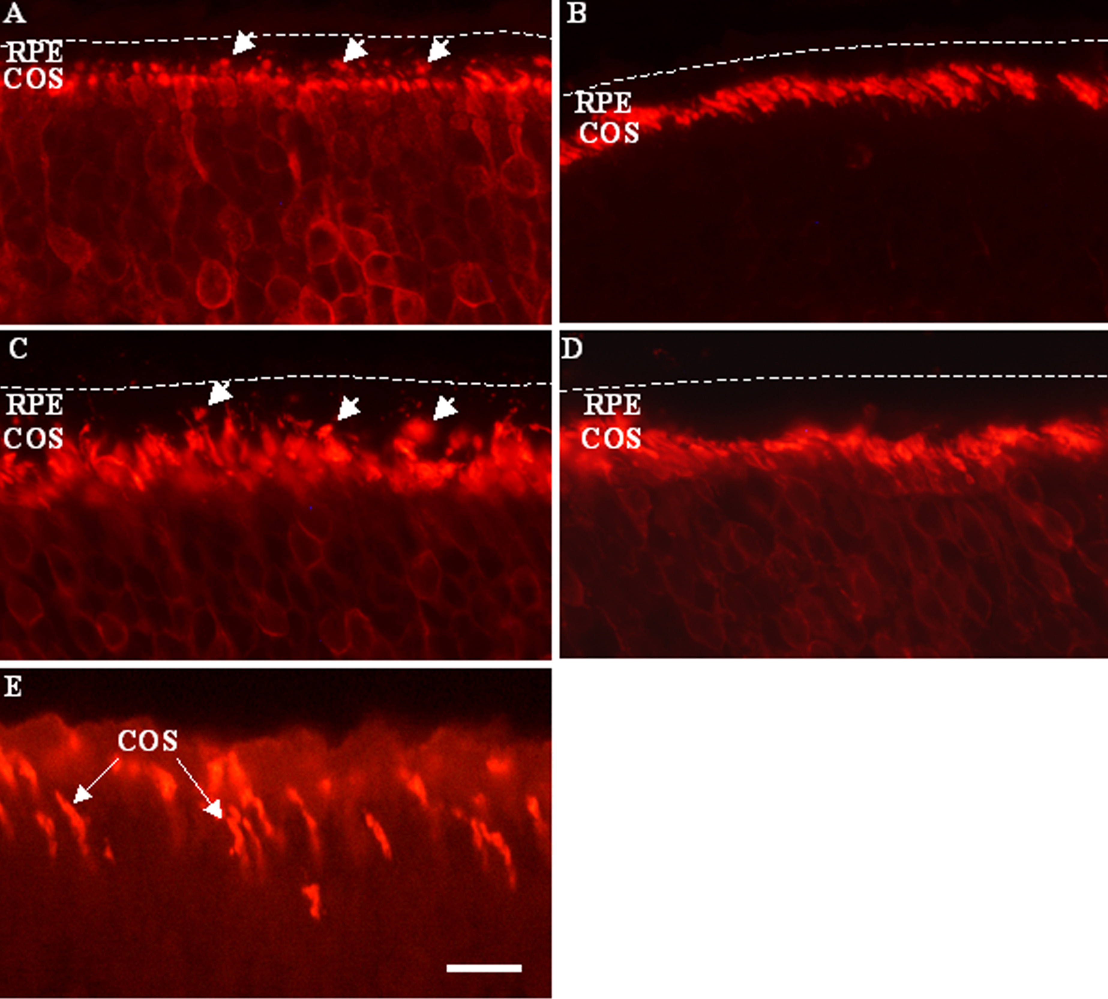
 Figure 2 of Krigel, Mol Vis 2010; 16:2873-2881.