Figure 2. Expression of human retinal ganglion cell (RGC)-specific markers. A-F: Immunofluorescence of human RGCs at day 3 of culture shows positive expression of Thy-1 (B) and TUJ1 (E). DAPI nuclear staining is shown in (A) and (D) and in the merged images in (C) and (F). Confocal double immunofluorescence shows the co-staining of Brn3a (G) and Thy1 (H) in cells at day 3 of culture (merged image in I), and Brn3a (J) and TUJI (K) in cells at day 7 (merged image in L). Scale bars: 25 µm.
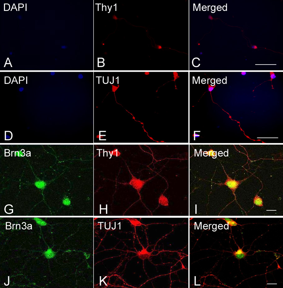
 Figure 2 of
Zhang, Mol Vis 2010; 16:2867-2872.
Figure 2 of
Zhang, Mol Vis 2010; 16:2867-2872.