Figure 1. Isolated human retinal ganglion cells (RGCs) in culture. A-D: Morphological changes of human RGCs in serum-free defined culture at different time intervals. The cell bodies and neurites
were examined by calcein-AM staining. (A) day 1; (B) day 7; (C) day 21 at lower magnification; (D) day 21 at higher magnification. E: Percentage of surviving RGCs over time. Approximately 50% of the RGCs survived after the first 3 days in culture. The survival
percentages were moderately reduced and maintained at about 20% after 1 month. Experiments were performed in triplicate. Error
bars: SD. Scale bars: (A, B, D) 50 µm, (C) 25 µm.
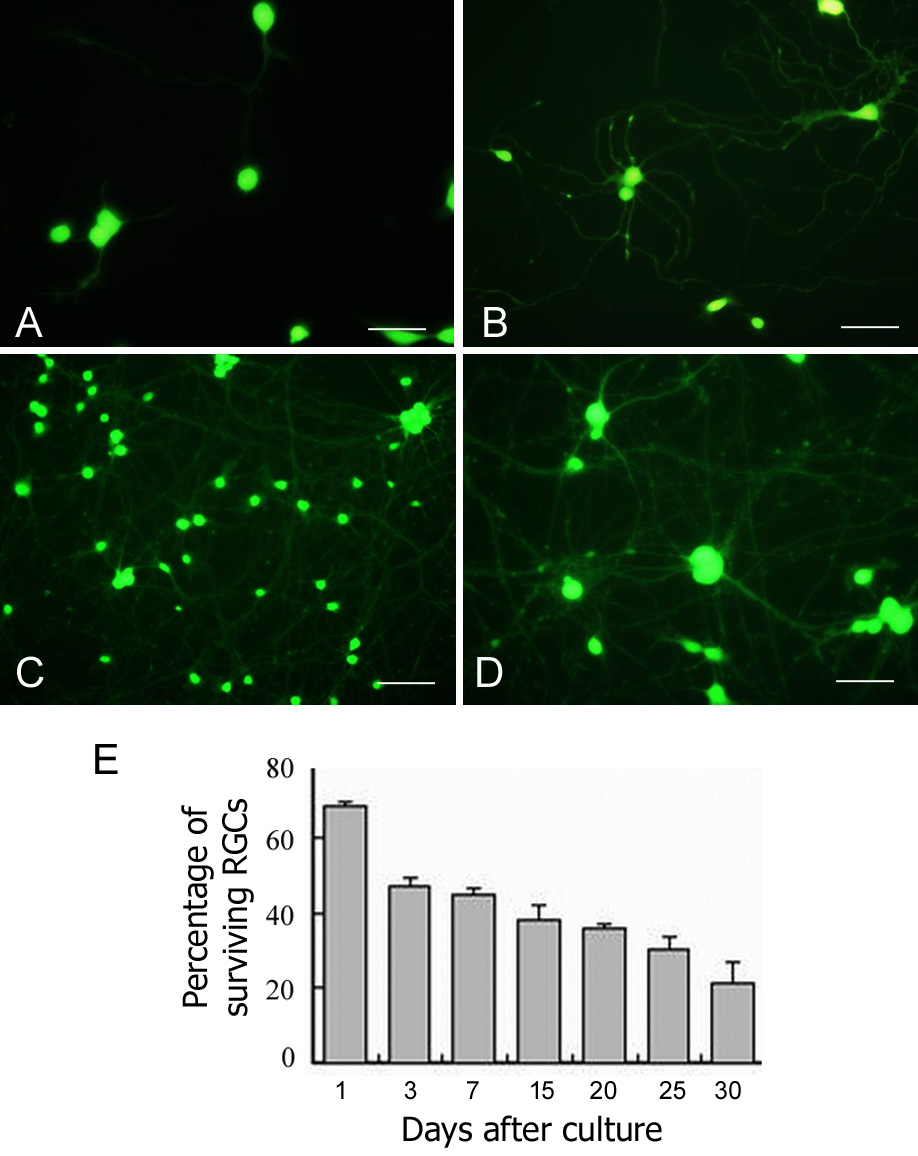
 Figure 1 of
Zhang, Mol Vis 2010; 16:2867-2872.
Figure 1 of
Zhang, Mol Vis 2010; 16:2867-2872.