Figure 2. Changes in retinal progenitor
cell morphology under different culture conditions. Retinal progenitor
cell s were cultured in the same serum-free base media, but under four
different treatment conditions defined by the presence or absence of
added growth factors, as follows: 1) no growth factor (A-C),
2)
glial cell line-derived neurotrophic factor (GDNF) alone (D-F),
3)
epidermal growth factor (EGF) alone (G-I), and 4)
EGF+GDNF (J-L). In each case, EGF was used at a final
concentration of 20 ng/ml and GDNF at 10 ng/ml. The morphology of cells
in each condition was assessed on day 1, 3, and 5. Increased extension
of processes appeared in the “no growth factor” group (A-C;
D-F), with similar changes observed in the “GDNF alone”
group (A-C; D-F). Cells grown in EGF+GDNF
appeared to form more and larger spherical cellular aggregates
(spheres) over the course of 5 days. Magnification was ×100. Scale
bars: 100 μm.
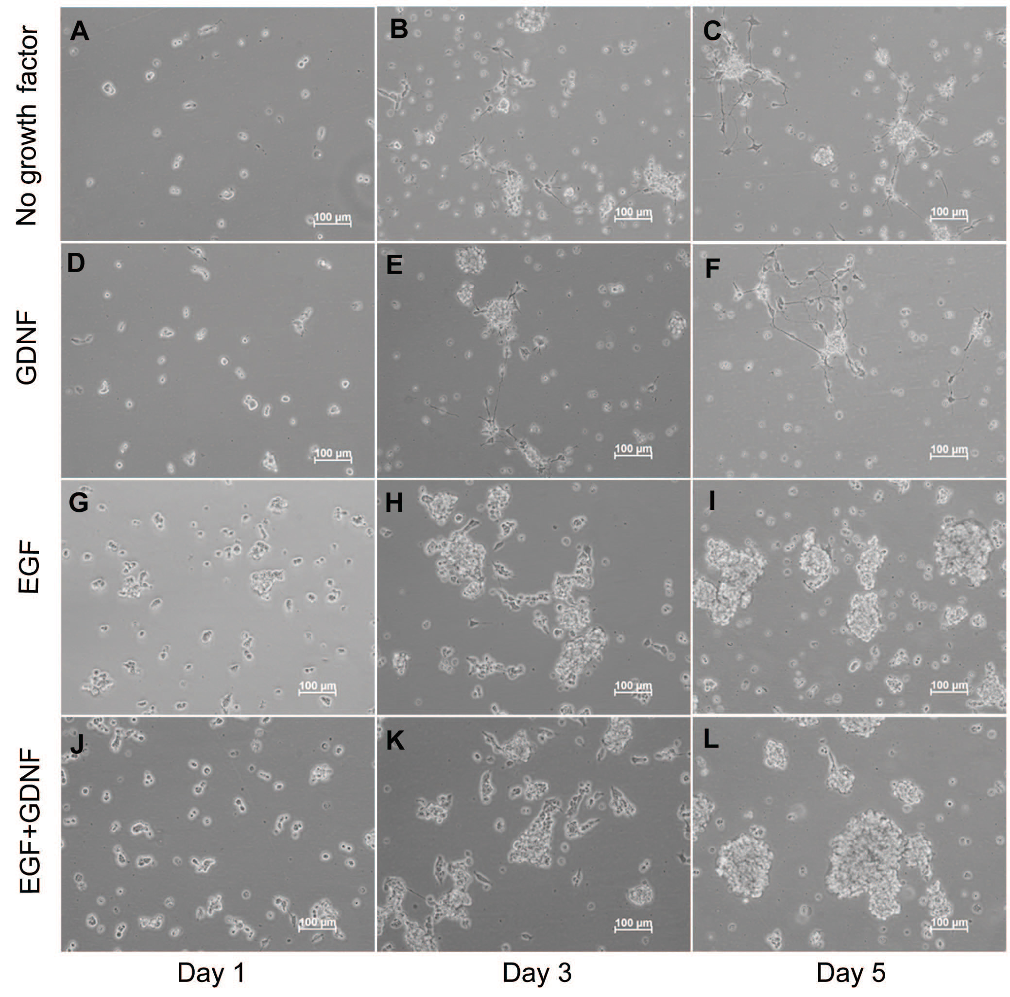
 Figure 2 of Wang, Mol Vis 2010; 16:2850-2866.