Figure 3 of
Duan, Mol Vis 2010; 16:2839-2846.
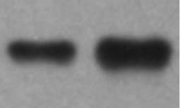
Figure 3.
Western blotting analysis of PGDS in AH. The left lane is a sample from a control and the right lane is a sample from a POAG patient.
 Figure 3 of
Duan, Mol Vis 2010; 16:2839-2846. Figure 3 of
Duan, Mol Vis 2010; 16:2839-2846.
Figure 3 of
Duan, Mol Vis 2010; 16:2839-2846. Figure 3 of
Duan, Mol Vis 2010; 16:2839-2846.