Figure 4. Confocal microscopic images of
HLE-3B and HeLa cells transfected with His- tagged pcDNA(3.1+)γDWT and
pcDNA(3.1+)γDE107A constructs. Wild-type and mutant proteins were
probed with anti-his antibody (raised in mouse), and FITC conjugated
anti-mouse secondary antibody. A, D, G, J:
This
panel set represents the protein visualized using FITC alone. B,
E, H, K: This panel set is a visualization of
the cells using propidium iodide as the nuclear dye. C, F,
I, L: This panel set shows merged signals. Notice that
the mutant shows punctate particles in both HLE-3B and HeLa cells,
while the wild-type molecule does not. The signals were magnified 630×.
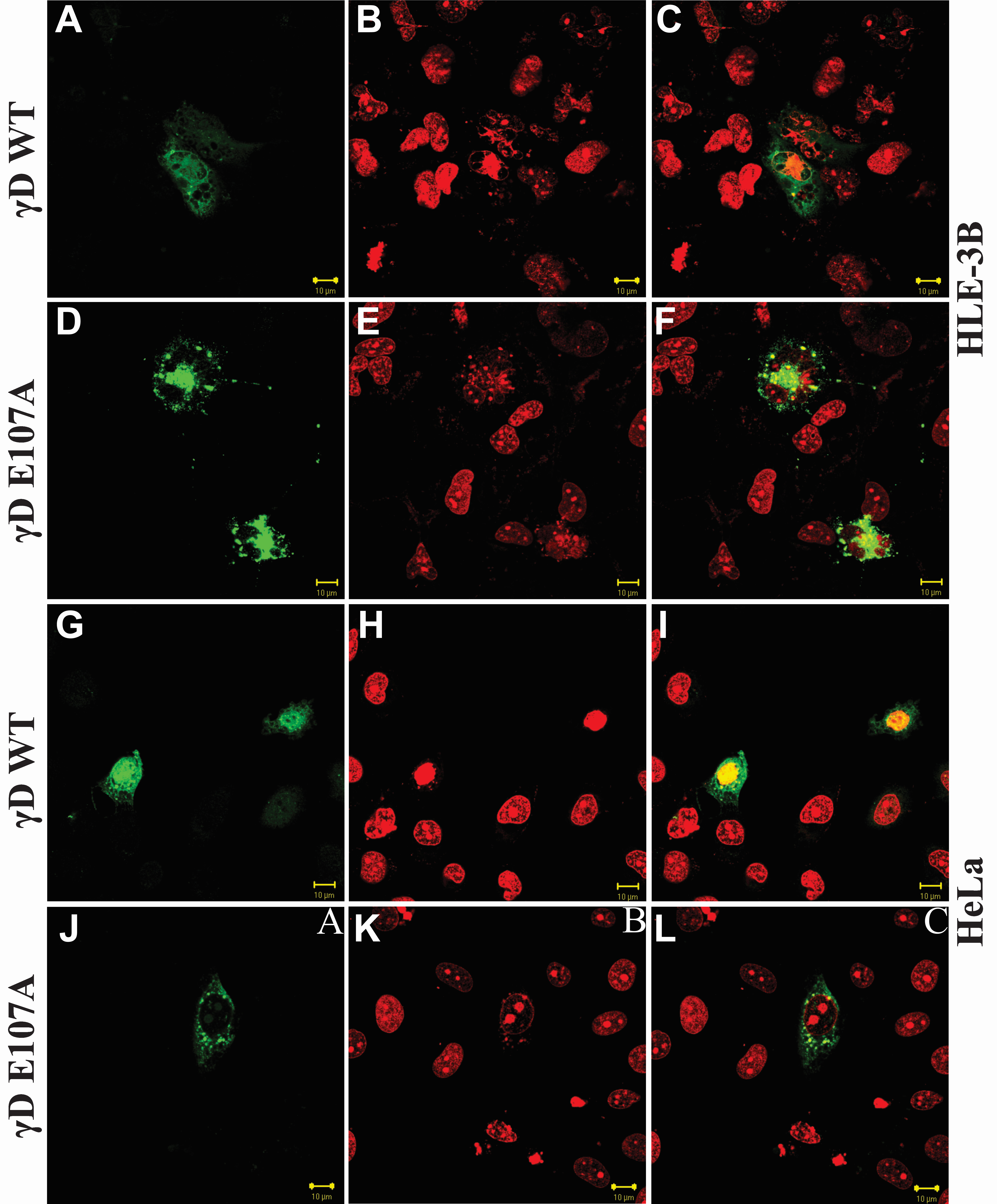
 Figure 4 of Vendra, Mol Vis 2010; 16:2822-2828.