Figure 1. Multifocal retinal dysplasia and canine multifocal retinopathy (cmr) fundus phenotypes. Fundus appearance associated with multifocal retinal dysplasia (MRD; A, B) compared to canine multifocal retinopathy 3 (cmr3; C, D), canine multifocal retinopathy 1 (cmr1; E), and a normal reference fundus (F). A-B: MRD: retinal folds with hyporeflective areas. Smaller lesions (arrows) can appear similar to cmr, particularly in dogs that have only one or few folds. C-D:cmr3: Multiple brown-gray oval lesions located subretinally (C, arrows) that generally are of smaller diameter than the optic disc. Lesions are elevated and are surrounded by a “halo”
of presumably clear subretinal fluid (D, arrow). E. Lesions typical for cmr1 (arrows). F: Normal fundus appearance.
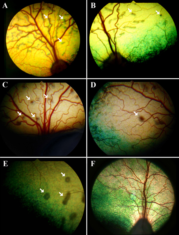
 Figure 1 of
Zangerl, Mol Vis 2010; 16:2791-2804.
Figure 1 of
Zangerl, Mol Vis 2010; 16:2791-2804.