Figure 7. The size exclusion chromatograms of HGDC samples with and without UV-C irradiation. All protein samples were denatured by
mixing 2% SDS and 10 mM β-mercaptoethanol (β-ME). The collected HGDC samples were first passed through 0.22 μm filter units,
and then subjected to size exclusion chromatography (SEC) analysis. The operating conditions of SEC analysis used were: injection
volume, 1 ml; flow rate of mobile phase, 0.25 ml/min; detector, UV at 280 nm; column, Superdex™ 75 10/300GL (GE Healthcare);
mobile phase composition, 5 mM sodium phosphate, 2% SDS, 10 mM β-ME (pH 7.0). Chromatogram 1: HGDC without UV-C irradiation
(exposure time=0); chromatogram 2: HGDC with 30 min-UV-C irradiation; chromatogram 3: HGDC with 60 min-UV-C irradiation. The
peak that was observed to elute at ~91 min was verified to correspond to β-ME according to the control experiment (chromatogram
of β-ME alone not shown).
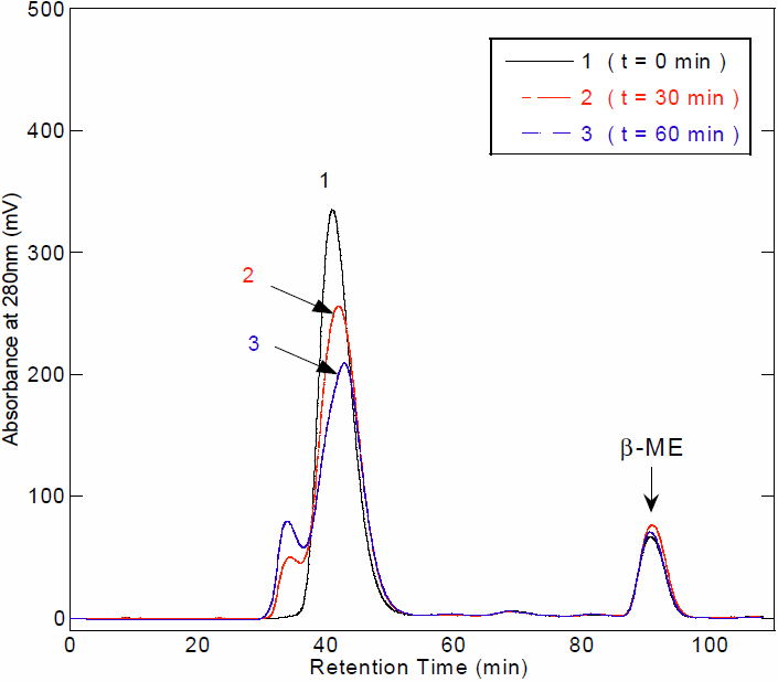
 Figure 7 of
Wang, Mol Vis 2010; 16:2777-2790.
Figure 7 of
Wang, Mol Vis 2010; 16:2777-2790.