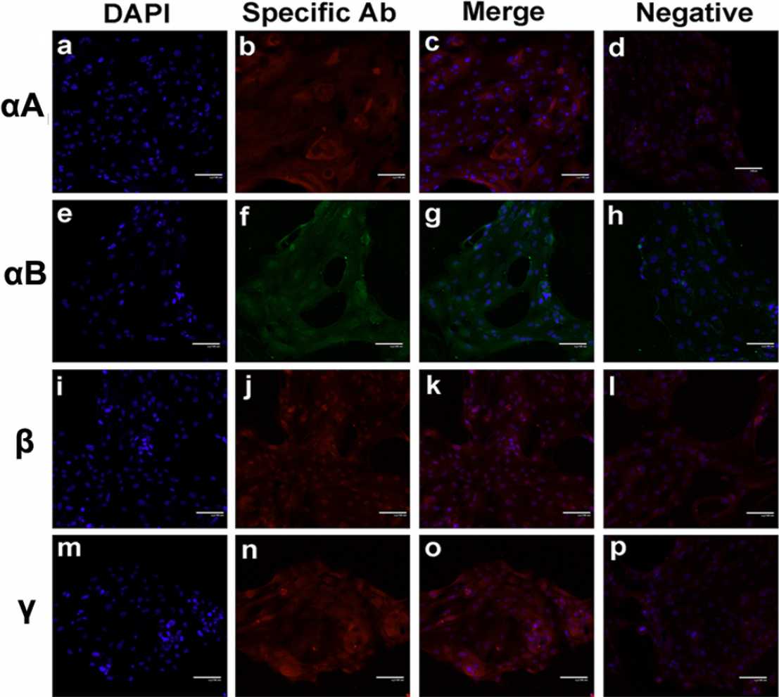
Figure 4. Lens crystallins expression in primary culture of human fetal corneal epithelial cell culture. Intact corneas from miscarried
fetus were used for culture of primary corneal epithelial cells, and second passages were used for immunofluorescence staining
for the proteins of αA-,αB-,β- and γ-crystallin using different combination of primary and fluorescence conjugated secondary
antibodies. The nuclei were stained with DAPI. Each section was observed under appropriate filters and the image of nuclei
was overlaid on image of protein staining (“merge”), respectively. For background controlling, the primary antibodies were
omitted in parallel protocol but only stained with secondary antibodies (“negative”).

 Figure 4 of
Ren, Mol Vis 2010; 16:2745-2752.
  Figure 4 of
Ren, Mol Vis 2010; 16:2745-2752.