Figure 5. Immunofluorescence staining of
mucin-5AC in the limbal epithelial cells treated with 100 ng/ml nerve
growth factor. A few of goblet cells (green, white arrows) were
detected at 24 h (A), the mumber of goblet cells increased at
the time point of 5 d comparing with 24 h (D; p<0.05). Nuclei
were stained with 4,6-diamidino-2-phenylindole (B, E;
blue). Double immunolabeling for mucin-5AC and
4,6-diamidino-2-phenylindole indicated the presence of goblet cells in
primary culture of mouse limbal epithelial cells with 100 ng/ml nerve
growth factor (C, F).
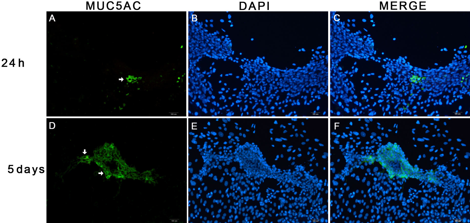
 Figure 5 of Li, Mol Vis 2010; 16:2739-2744.