Figure 4. Immunofluorescence staining of mucin-5AC in the control group (0 ng/ml nerve growth factor) at the time point of 5 d. The
results showed that mucin-5AC positive cells (goblet cells) were not detected when cultured for 5 d (A). Nuclei were stained with 4,6-diamidino-2-phenylindole (B; blue).
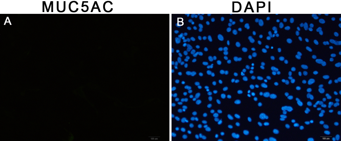
 Figure 4 of
Li, Mol Vis 2010; 16:2739-2744.
Figure 4 of
Li, Mol Vis 2010; 16:2739-2744.