Figure 1 of
Li, Mol Vis 2010; 16:2739-2744.
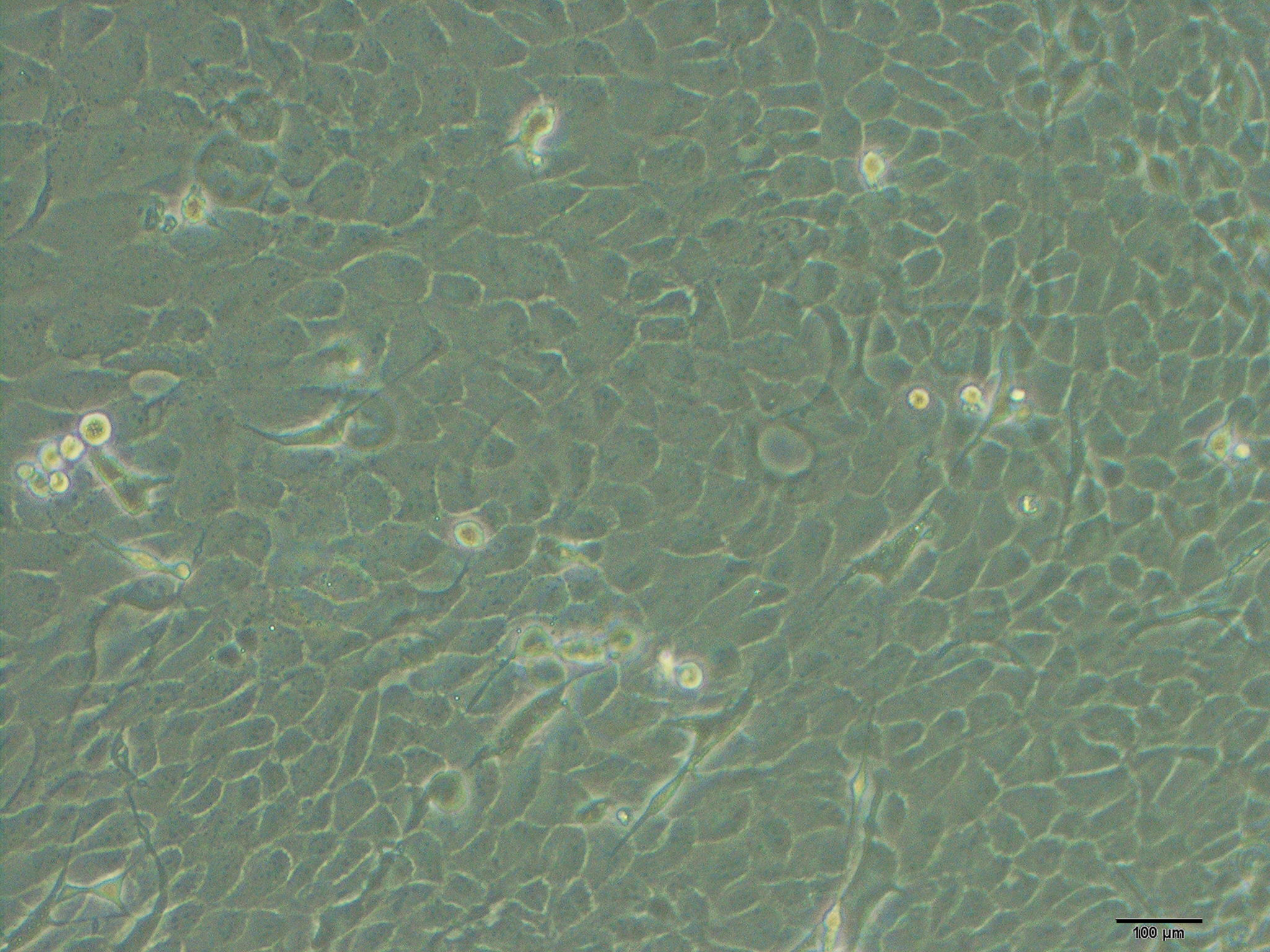
Figure 1.
Characterization of primary culture of mouse limbal epithelial cells. Phase contrast microscopy showed the morphology of limbal epithelial cells. Cells appeared to be compact, uniform and cobblestone pavement in shape.
 Figure 1 of Li, Mol Vis 2010; 16:2739-2744.