Figure 1. Representative immunostaining results for β-catenin and cyclin D1. A: the negative control has the first antibody replaced with IgG. B: β-catenin protein expression detected in the membrane (400×), C, aberrant localization of β-catenin in the cytoplasm/nuclei (400×), and D, cyclin D1 protein expression in nuclei (400×).
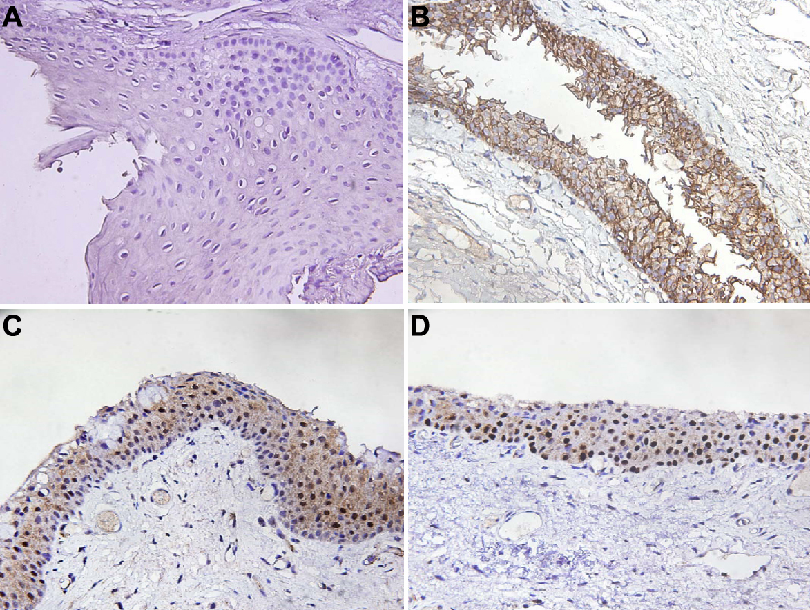
 Figure 1 of
Tung, Mol Vis 2010; 16:2733-2738.
Figure 1 of
Tung, Mol Vis 2010; 16:2733-2738.