Figure 6. Immunostaining of cytospin of pannus tissue. Double-immunostaining for p63 (red) and α-SMA (green) was observed in individual
cells (indicated by arrows) in cytospin of pannus (A, with DAPI staining; B, without DAPI staining). Scale bar indicates 50 μm.
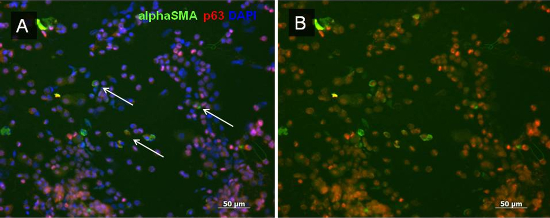
 Figure 6 of
Kawashima, Mol Vis 2010; 16:2727-2732.
Figure 6 of
Kawashima, Mol Vis 2010; 16:2727-2732.