Figure 5. Immunostaining of pannus tissue against p63 and α-SMA. Nuclear p63 (red) and cytoplasmic α- SMA (green) double-positive cells
were observed in subepithelial location in pannus tissue (A and B). B was syntheses photo of p63 (C), α- SMA (D), and DAPI (E). Scale bar indicates 50 μm.
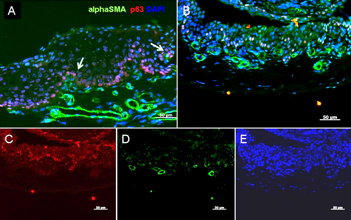
 Figure 5 of
Kawashima, Mol Vis 2010; 16:2727-2732.
Figure 5 of
Kawashima, Mol Vis 2010; 16:2727-2732.