Figure 4. Immunohistochemistry of E-cadherin and β-catenin. Epithelial basal cells in pannus showed weak staining for E-cadherin (green)
and translocation of β-catenin (red) from intercellular junctions to cytoplasm. A: Pannus with double-staining for E-cadherin and β-catenin. B: Normal cornea. C: βcatenin, D: E-cadherin, E: E-cadherin and DAPI, in pannus epithelium was shown. A was a combination of C, D, and E. The scale bar indicates 50 μm.
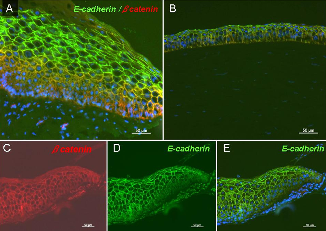
 Figure 4 of
Kawashima, Mol Vis 2010; 16:2727-2732.
Figure 4 of
Kawashima, Mol Vis 2010; 16:2727-2732.